2BGV

X-ray structure of ferric cytochrome c-550 from Paracoccus versutus


Experimental Data Snapshot

  • Method: X-RAY DIFFRACTION
  • Resolution: 1.90 Å
  • R-Value Free: 0.191 
  • R-Value Work: 0.158 
  • R-Value Observed: 0.160 

wwPDB Validation   3D Report Full Report


Ligand Structure Quality Assessment 


This is version 1.2 of the entry. See complete history


Literature

The Effect of Replacing the Axial Methionine Ligand with a Lysine Residue in Cytochrome C-550 from Paracoccus Versutus Assessed by X-Ray Crystallography and Unfolding.

Worrall, J.A.R.Van Roon, A.-M.M.Ubbink, M.Canters, G.W.

(2005) FEBS J 272: 2441

  • DOI: https://doi.org/10.1111/j.1742-4658.2005.04664.x
  • Primary Citation of Related Structures:  
    2BGV, 2BH4, 2BH5

  • PubMed Abstract: 

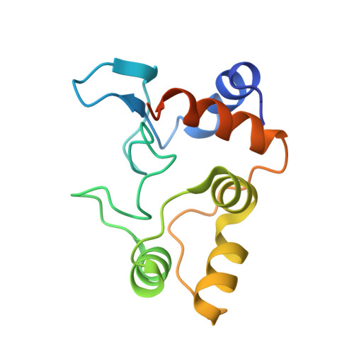
    The structure of cytochrome c-550 from the nonphotosynthetic bacteria Paraccocus versutus has been solved by X-ray crystallography to 1.90 A resolution, and reveals a high structural homology to other bacterial cytochromes c(2). The effect of replacing the axial heme-iron methionine ligand with a lysine residue on protein structure and unfolding has been assessed using the M100K variant. From X-ray structures at 1.95 and 1.55 A resolution it became clear that the amino group of the lysine side chain coordinates to the heme-iron. Structural differences compared to the wild-type protein are confined to the lysine ligand loop connecting helices four and five. In the heme cavity an additional water molecule is found which participates in an H-bonding interaction with the lysine ligand. Under cryo-conditions extra electron density in the lysine ligand loop is revealed, leading to residues K97 to T101 being modeled with a double main-chain conformation. Upon unfolding, dissociation of the lysine ligand from the heme-iron is shown to be pH dependent, with NMR data consistent with the occurrence of a ligand exchange mechanism similar to that seen for the wild-type protein.


  • Organizational Affiliation

    Leiden Institute of Chemistry, Leiden University, Gorlaeus Laboratories, Leiden, the Netherlands.


Macromolecules
Find similar proteins by:  (by identity cutoff)  |  3D Structure
Entity ID: 1
MoleculeChains Sequence LengthOrganismDetailsImage
CYTOCHROME C-550A [auth X]134Paracoccus versutusMutation(s): 0 
UniProt
Find proteins for Q00499 (Paracoccus versutus)
Explore Q00499 
Go to UniProtKB:  Q00499
Entity Groups  
Sequence Clusters30% Identity50% Identity70% Identity90% Identity95% Identity100% Identity
UniProt GroupQ00499
Sequence Annotations
Expand
  • Reference Sequence
Small Molecules
Ligands 1 Unique
IDChains Name / Formula / InChI Key2D Diagram3D Interactions
HEC
Query on HEC

Download Ideal Coordinates CCD File 
B [auth X]HEME C
C34 H34 Fe N4 O4
HXQIYSLZKNYNMH-LJNAALQVSA-N
Experimental Data & Validation

Experimental Data

  • Method: X-RAY DIFFRACTION
  • Resolution: 1.90 Å
  • R-Value Free: 0.191 
  • R-Value Work: 0.158 
  • R-Value Observed: 0.160 
  • Space Group: P 41 21 2
Unit Cell:
Length ( Å )Angle ( ˚ )
a = 57.102α = 90
b = 57.102β = 90
c = 66.616γ = 90
Software Package:
Software NamePurpose
REFMACrefinement
HKL-2000data reduction
SCALEPACKdata scaling
MOLREPphasing

Structure Validation

View Full Validation Report


Ligand Structure Quality Assessment 


Entry History 

Deposition Data

Revision History  (Full details and data files)

  • Version 1.0: 2005-05-11
    Type: Initial release
  • Version 1.1: 2011-05-08
    Changes: Version format compliance
  • Version 1.2: 2011-07-13
    Changes: Version format compliance