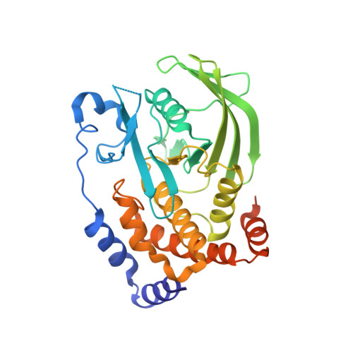
2BGD | pdb_00002bgd

Structure-based design of Protein Tyrosine Phosphatase-1B Inhibitors


Experimental Data Snapshot

  • Method: X-RAY DIFFRACTION
  • Resolution: 2.40 Å
  • R-Value Free: 
    0.200 (Depositor), 0.208 (DCC) 
  • R-Value Work: 
    0.164 (Depositor), 0.177 (DCC) 
  • R-Value Observed: 
    0.166 (Depositor) 

wwPDB Validation 3D Report Full Report

Validation slider image for 2BGD

Ligand Structure Quality Assessment 


This is version 1.4 of the entry. See complete history. 

Literature

Structure-Based Design of Protein Tyrosine Phosphatase-1B Inhibitors

Black, E., Breed, J., Breeze, A.L., Embrey, K., Garcia, R., Gero, T.W., Godfrey, L., Kenny, P.W., Morley, A.D., Minshull, C.A., Pannifer, A.D., Read, J., Rees, A., Russell, D.J., Toader, D., Tucker, J.

(2005) Bioorg Med Chem Lett 15: 2503

  • DOI: https://doi.org/10.1016/j.bmcl.2005.03.068
  • Primary Citation Related Structures: 
    2BGD, 2BGE

  • PubMed Abstract: 

    Using structure-based design, a new class of inhibitors of protein tyrosine phosphatase-1B (PTP1B) has been identified, which incorporate the 1,2,5-thiadiazolidin-3-one-1,1-dioxide template.


  • Organizational Affiliation: 
    • AstraZeneca, Mereside, Alderley Park, Macclesfield, Cheshire SK10 4TG, UK.

Macromolecule Content 

  • Total Structure Weight: 37.91 kDa 
  • Atom Count: 2,603 
  • Modeled Residue Count: 295 
  • Deposited Residue Count: 321 
  • Unique protein chains: 1

Macromolecules

Find similar proteins by:|  3D Structure
Entity ID: 1
MoleculeChains  Sequence LengthOrganismDetailsImage
PROTEIN-TYROSINE PHOSPHATASE NON-RECEPTOR TYPE 1321Homo sapiensMutation(s): 0 
EC: 3.1.3.48
UniProt & NIH Common Fund Data Resources
Find proteins for P18031 (Homo sapiens)
Explore P18031 
Go to UniProtKB:  P18031
PHAROS:  P18031
GTEx:  ENSG00000196396 
Entity Groups
Sequence Clusters30% Identity50% Identity70% Identity90% Identity95% Identity100% Identity
UniProt GroupP18031
Sequence Annotations
Expand
Reference Sequence

Small Molecules

Ligands 4 Unique
IDChains Name / Formula / InChI Key2D Diagram3D Interactions
T1D

Query on T1D


Download:Ideal Coordinates CCD File
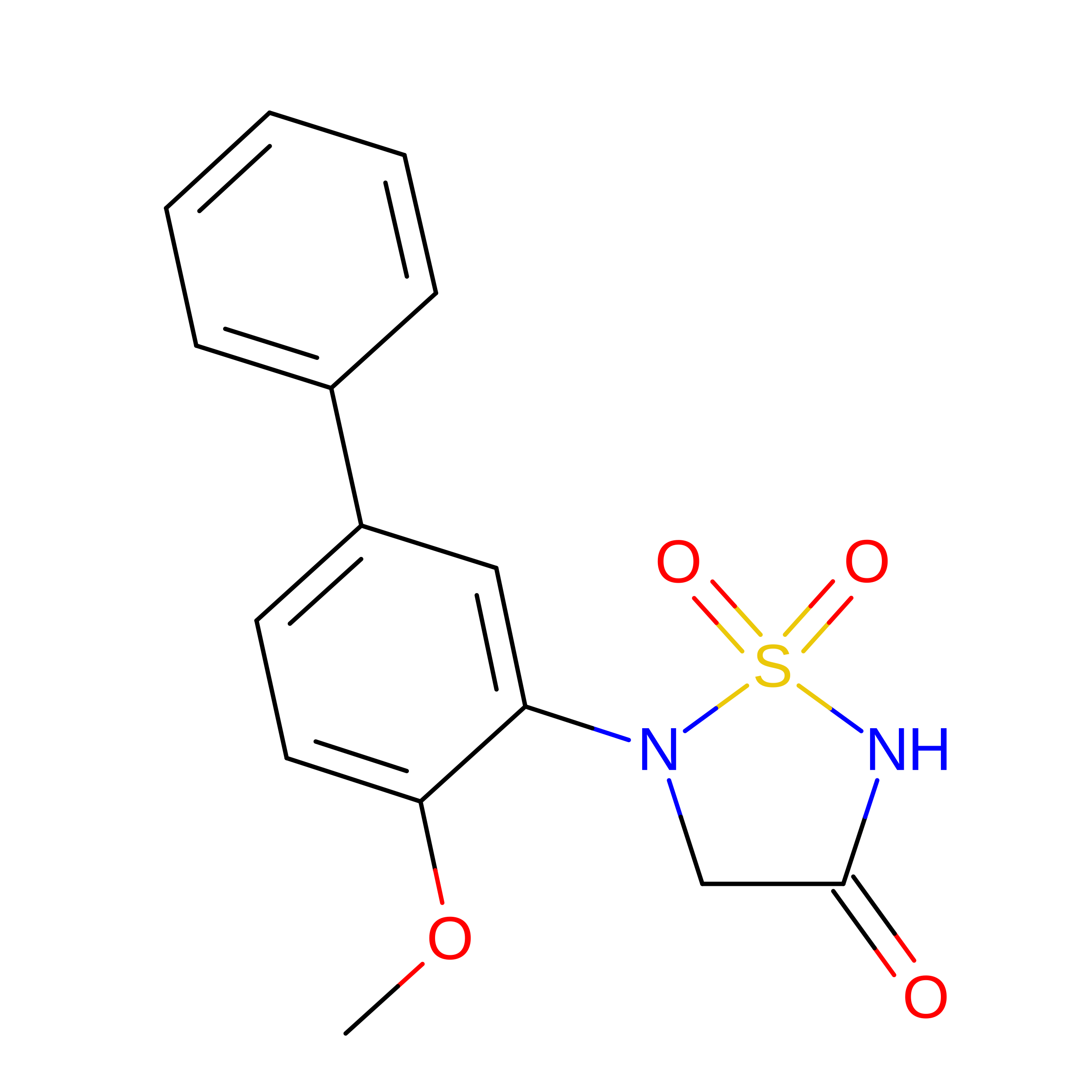
B [auth A]5-(4-METHOXYBIPHENYL-3-YL)-1,2,5-THIADIAZOLIDIN-3-ONE 1,1-DIOXIDE
C15 H14 N2 O4 S
IGXKSKWHHHYBFM-UHFFFAOYSA-N
PO4

Query on PO4


Download:Ideal Coordinates CCD File
F [auth A]PHOSPHATE ION
O4 P
NBIIXXVUZAFLBC-UHFFFAOYSA-K
CL

Query on CL


Download:Ideal Coordinates CCD File
C [auth A],
D [auth A],
E [auth A]
CHLORIDE ION
Cl
VEXZGXHMUGYJMC-UHFFFAOYSA-M
NA

Query on NA


Download:Ideal Coordinates CCD File
G [auth A]SODIUM ION
Na
FKNQFGJONOIPTF-UHFFFAOYSA-N

Experimental Data & Validation

Experimental Data

  • Method: X-RAY DIFFRACTION
  • Resolution: 2.40 Å
  • R-Value Free:  0.200 (Depositor), 0.208 (DCC) 
  • R-Value Work:  0.164 (Depositor), 0.177 (DCC) 
  • R-Value Observed: 0.166 (Depositor) 
Space Group: P 31 2 1
Unit Cell:
Length ( Å )Angle ( ˚ )
a = 88.908α = 90
b = 88.908β = 90
c = 104.548γ = 120
Software Package:
Software NamePurpose
REFMACrefinement
MOSFLMdata reduction
SCALAdata scaling
AMoREphasing

Structure Validation

View Full Validation Report


Ligand Structure Quality Assessment 


Entry History 

Revision History  (Full details and data files)

  • Version 1.0: 2005-05-04
    Type: Initial release
  • Version 1.1: 2011-07-13
    Changes: Advisory, Refinement description, Version format compliance
  • Version 1.2: 2015-09-16
    Changes: Source and taxonomy
  • Version 1.3: 2019-07-24
    Changes: Data collection
  • Version 1.4: 2024-05-08
    Changes: Data collection, Database references, Derived calculations, Other