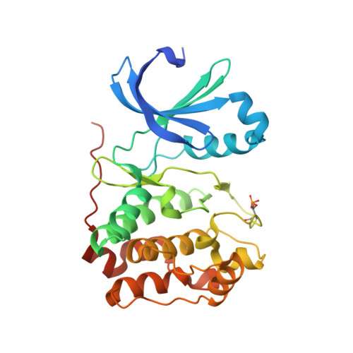
2BFY

Complex of Aurora-B with INCENP and Hesperadin.


Experimental Data Snapshot

  • Method: X-RAY DIFFRACTION
  • Resolution: 1.80 Å
  • R-Value Free: 0.226 
  • R-Value Work: 0.203 
  • R-Value Observed: 0.203 

wwPDB Validation   3D Report Full Report


Ligand Structure Quality Assessment 


This is version 1.3 of the entry. See complete history


Literature

Mechanism of Aurora B Activation by Incenp and Inhibition by Hesperadin

Sessa, F.Mapelli, M.Ciferri, C.Tarricone, C.Areces, L.B.Schneider, T.R.Stukenberg, P.T.Musacchio, A.

(2005) Mol Cell 18: 379

  • DOI: https://doi.org/10.1016/j.molcel.2005.03.031
  • Primary Citation of Related Structures:  
    2BFX, 2BFY

  • PubMed Abstract: 

    Aurora family serine/threonine kinases control mitotic progression, and their deregulation is implicated in tumorigenesis. Aurora A and Aurora B, the best-characterized members of mammalian Aurora kinases, are approximately 60% identical but bind to unrelated activating subunits. The structure of the complex of Aurora A with the TPX2 activator has been reported previously. Here, we report the crystal structure of Aurora B in complex with the IN-box segment of the inner centromere protein (INCENP) activator and with the small molecule inhibitor Hesperadin. The Aurora B:INCENP complex is remarkably different from the Aurora A:TPX2 complex. INCENP forms a crown around the small lobe of Aurora B and induces the active conformation of the T loop allosterically. The structure represents an intermediate state of activation of Aurora B in which the Aurora B C-terminal segment stabilizes an open conformation of the catalytic cleft, and a critical ion pair in the kinase active site is impaired. Phosphorylation of two serines in the carboxyl terminus of INCENP generates the fully active kinase.


  • Organizational Affiliation

    Department of Experimental Oncology, European Institute of Oncology, Via Ripamonti 435, 20141 Milan, Italy.


Macromolecules
Find similar proteins by:  (by identity cutoff)  |  3D Structure
Entity ID: 1
MoleculeChains Sequence LengthOrganismDetailsImage
AURORA KINASE B-A
A, B
284Xenopus laevisMutation(s): 1 
EC: 2.7.11.1
UniProt
Find proteins for Q6DE08 (Xenopus laevis)
Explore Q6DE08 
Go to UniProtKB:  Q6DE08
Entity Groups  
Sequence Clusters30% Identity50% Identity70% Identity90% Identity95% Identity100% Identity
UniProt GroupQ6DE08
Sequence Annotations
Expand
  • Reference Sequence
Find similar proteins by:  (by identity cutoff)  |  3D Structure
Entity ID: 2
MoleculeChains Sequence LengthOrganismDetailsImage
INNER CENTROMERE PROTEIN A
C, D
43Xenopus laevisMutation(s): 0 
UniProt
Find proteins for O13024 (Xenopus laevis)
Explore O13024 
Go to UniProtKB:  O13024
Entity Groups  
Sequence Clusters30% Identity50% Identity70% Identity90% Identity95% Identity100% Identity
UniProt GroupO13024
Sequence Annotations
Expand
  • Reference Sequence
Small Molecules
Ligands 1 Unique
IDChains Name / Formula / InChI Key2D Diagram3D Interactions
H1N
Query on H1N

Download Ideal Coordinates CCD File 
E [auth A],
F [auth B]
N-[2-OXO-3-((E)-PHENYL{[4-(PIPERIDIN-1-YLMETHYL)PHENYL]IMINO}METHYL)-2,6-DIHYDRO-1H-INDOL-5-YL]ETHANESULFONAMIDE
C29 H32 N4 O3 S
SGZZQKMOFHIDKW-MCKMWFOCSA-N
Modified Residues  1 Unique
IDChains TypeFormula2D DiagramParent
TPO
Query on TPO
A, B
L-PEPTIDE LINKINGC4 H10 N O6 PTHR
Binding Affinity Annotations 
IDSourceBinding Affinity
H1N BindingDB:  2BFY IC50: 3 (nM) from 1 assay(s)
Experimental Data & Validation

Experimental Data

  • Method: X-RAY DIFFRACTION
  • Resolution: 1.80 Å
  • R-Value Free: 0.226 
  • R-Value Work: 0.203 
  • R-Value Observed: 0.203 
  • Space Group: P 1 21 1
Unit Cell:
Length ( Å )Angle ( ˚ )
a = 45.945α = 90
b = 67.044β = 96.51
c = 116.462γ = 90
Software Package:
Software NamePurpose
CNSrefinement
HKL-2000data reduction
SCALEPACKdata scaling
AMoREphasing

Structure Validation

View Full Validation Report


Ligand Structure Quality Assessment 


Entry History 

Deposition Data

Revision History  (Full details and data files)

  • Version 1.0: 2005-05-03
    Type: Initial release
  • Version 1.1: 2014-05-14
    Changes: Database references, Derived calculations, Non-polymer description, Other, Refinement description, Source and taxonomy, Structure summary, Version format compliance
  • Version 1.2: 2015-05-20
    Changes: Structure summary
  • Version 1.3: 2023-12-13
    Changes: Data collection, Database references, Derived calculations, Other, Refinement description