Bacterial Chromosome Segregation: Structure and DNA Binding of the Soj Dimer--A Conserved Biological Switch
Leonard, T.A., Butler, P.J.G., Lowe, J.(2005) EMBO J 24: 270
- PubMed: 15635448 Search on PubMedSearch on PubMed Central
- DOI: https://doi.org/10.1038/sj.emboj.7600530
- Primary Citation Related Structures:
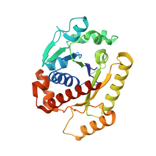
1WCV, 2BEJ, 2BEK - PubMed Abstract:
Soj and Spo0J of the Gram-negative hyperthermophile Thermus thermophilus belong to the conserved ParAB family of bacterial proteins implicated in plasmid and chromosome partitioning. Spo0J binds to DNA near the replication origin and localises at the poles following initiation of replication. Soj oscillates in the nucleoid region in an ATP- and Spo0J-dependent fashion. Here, we show that Soj undergoes ATP-dependent dimerisation in solution and forms nucleoprotein filaments with DNA. Crystal structures of Soj in three nucleotide states demonstrate that the empty and ADP-bound states are monomeric, while a hydrolysis-deficient mutant, D44A, is capable of forming a nucleotide 'sandwich' dimer. Soj ATPase activity is stimulated by Spo0J or the N-terminal 20 amino-acid peptide of Spo0J. Our analysis shows that dimerisation and activation involving a peptide containing a Lys/Arg is conserved for Soj, ParA and MinD and their modulators Spo0J, ParB and MinE, respectively. By homology to the nitrogenase iron protein and the GTPases Ffh/FtsY, we suggest that Soj dimerisation and regulation represent a conserved biological switch.
- MRC Laboratory of Molecular Biology, Cambridge, UK. tleonard@mrc-lmb.cam.ac.uk
Organizational Affiliation: