2BCD

X-ray crystal structure of Protein Phosphatase-1 with the marine toxin motuporin bound


Experimental Data Snapshot

  • Method: X-RAY DIFFRACTION
  • Resolution: 2.10 Å
  • R-Value Free: 0.264 
  • R-Value Work: 0.221 
  • R-Value Observed: 0.223 

wwPDB Validation   3D Report Full Report


This is version 2.0 of the entry. See complete history


Literature

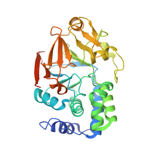
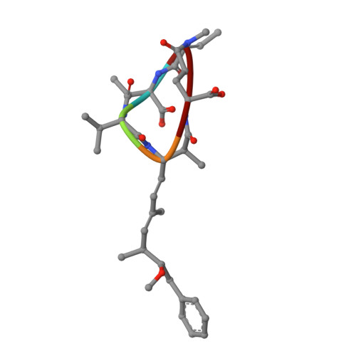
Crystal Structures of Protein Phosphatase-1 Bound to Motuporin and Dihydromicrocystin-LA: Elucidation of the Mechanism of Enzyme Inhibition by Cyanobacterial Toxins.

Maynes, J.T.Luu, H.A.Cherney, M.M.Andersen, R.J.Williams, D.Holmes, C.F.James, M.N.

(2006) J Mol Biol 356: 111-120

  • DOI: https://doi.org/10.1016/j.jmb.2005.11.019
  • Primary Citation of Related Structures:  
    2BCD, 2BDX

  • PubMed Abstract: 

    The microcystins and nodularins are tumour promoting hepatotoxins that are responsible for global adverse human health effects and wildlife fatalities in countries where drinking water supplies contain cyanobacteria. The toxins function by inhibiting broad specificity Ser/Thr protein phosphatases in the host cells, thereby disrupting signal transduction pathways. A previous crystal structure of a microcystin bound to the catalytic subunit of protein phosphatase-1 (PP-1c) showed distinct changes in the active site region when compared with protein phosphatase-1 structures bound to other toxins. We have elucidated the crystal structures of the cyanotoxins, motuporin (nodularin-V) and dihydromicrocystin-LA bound to human protein phosphatase-1c (gamma isoform). The atomic structures of these complexes reveal the structural basis for inhibition of protein phosphatases by these toxins. Comparisons of the structures of the cyanobacterial toxin:phosphatase complexes explain the biochemical mechanism by which microcystins but not nodularins permanently modify their protein phosphatase targets by covalent addition to an active site cysteine residue.


  • Organizational Affiliation

    Canadian Institutes of Health Research, Group in Protein Structure and Function Department of Biochemistry, Faculty of Medicine, University of Alberta, Edmonton, Alta, Canada T6G 2H7.


Macromolecules
Find similar proteins by:  (by identity cutoff)  |  3D Structure
Entity ID: 1
MoleculeChains Sequence LengthOrganismDetailsImage
Serine/threonine protein phosphatase PP1-gamma catalytic subunit323Homo sapiensMutation(s): 0 
Gene Names: PPP1CC
EC: 3.1.3.16
UniProt & NIH Common Fund Data Resources
Find proteins for P36873 (Homo sapiens)
Explore P36873 
Go to UniProtKB:  P36873
PHAROS:  P36873
GTEx:  ENSG00000186298 
Entity Groups  
Sequence Clusters30% Identity50% Identity70% Identity90% Identity95% Identity100% Identity
UniProt GroupP36873
Sequence Annotations
Expand
  • Reference Sequence

Find similar proteins by:  Sequence   |   3D Structure  

Entity ID: 2
MoleculeChains Sequence LengthOrganismDetailsImage
MOTUPORIN5Theonella sp.Mutation(s): 0 
Sequence Annotations
Expand
  • Reference Sequence
Small Molecules
Ligands 2 Unique
IDChains Name / Formula / InChI Key2D Diagram3D Interactions
BME
Query on BME

Download Ideal Coordinates CCD File 
C [auth A],
D [auth A]
BETA-MERCAPTOETHANOL
C2 H6 O S
DGVVWUTYPXICAM-UHFFFAOYSA-N
MN
Query on MN

Download Ideal Coordinates CCD File 
E [auth A]
F [auth A]
G [auth A]
H [auth A]
I [auth A]
E [auth A],
F [auth A],
G [auth A],
H [auth A],
I [auth A],
J [auth A],
K [auth A],
L [auth A]
MANGANESE (II) ION
Mn
WAEMQWOKJMHJLA-UHFFFAOYSA-N
Biologically Interesting Molecules (External Reference) 1 Unique
Experimental Data & Validation

Experimental Data

  • Method: X-RAY DIFFRACTION
  • Resolution: 2.10 Å
  • R-Value Free: 0.264 
  • R-Value Work: 0.221 
  • R-Value Observed: 0.223 
  • Space Group: P 42 21 2
Unit Cell:
Length ( Å )Angle ( ˚ )
a = 100.955α = 90
b = 100.955β = 90
c = 63.485γ = 90
Software Package:
Software NamePurpose
SCALAdata scaling
AMoREphasing
REFMACrefinement
PDB_EXTRACTdata extraction
MOSFLMdata reduction
CCP4data scaling

Structure Validation

View Full Validation Report


Entry History 

Deposition Data

Revision History  (Full details and data files)

  • Version 1.0: 2006-01-17
    Type: Initial release
  • Version 1.1: 2008-05-01
    Changes: Version format compliance
  • Version 1.2: 2011-07-13
    Changes: Atomic model, Database references, Derived calculations, Non-polymer description, Structure summary, Version format compliance
  • Version 1.3: 2011-07-27
    Changes: Database references
  • Version 1.4: 2012-12-12
    Changes: Other
  • Version 1.5: 2023-08-23
    Changes: Advisory, Data collection, Database references, Derived calculations, Refinement description
  • Version 2.0: 2023-11-15
    Changes: Atomic model, Data collection, Derived calculations