2BC3 | pdb_00002bc3

T7-tagged full-length streptavidin


Experimental Data Snapshot

  • Method: X-RAY DIFFRACTION
  • Resolution: 1.54 Å
  • R-Value Free: 
    0.243 (Depositor) 
  • R-Value Work: 
    0.175 (Depositor), 0.185 (DCC) 
  • R-Value Observed: 
    0.175 (Depositor) 

Starting Model: experimental
View more details

wwPDB Validation 3D Report Full Report

Validation slider image for 2BC3

This is version 1.4 of the entry. See complete history

Literature

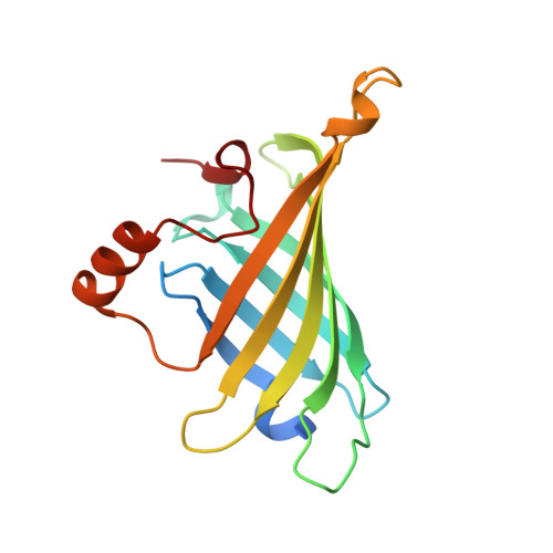
Crystallographic Analysis of a Full-length Streptavidin with Its C-terminal Polypeptide Bound in the Biotin Binding Site.

Le Trong, I.Humbert, N.Ward, T.R.Stenkamp, R.E.

(2006) J Mol Biology 356: 738-745

  • DOI: https://doi.org/10.1016/j.jmb.2005.11.086
  • Primary Citation Related Structures: 
    2BC3

  • PubMed Abstract: 

    The structure of a full-length streptavidin has been determined at 1.7 A resolution and shows that the 20 residue extension at the C terminus forms a well-ordered polypeptide loop on the surface of the tetramer. Residues 150-153 of the extension are bound to the ligand-binding site, possibly competing with exogenous ligands. The binding mode of these residues is compared with that of biotin and peptidic ligands. The observed structure helps to rationalize the observations that full-length mature streptavidin binds biotinylated macromolecules with reduced affinity.


  • Organizational Affiliation
    • Departments of Biological Structure and Biochemistry and the Biomolecular Structure Center, University of Washington, Box 357420, Seattle, WA 98195-7420, USA.

Macromolecule Content 

  • Total Structure Weight: 33.42 kDa 
  • Atom Count: 2,434 
  • Modeled Residue Count: 286 
  • Deposited Residue Count: 318 
  • Unique protein chains: 1

Macromolecules

Find similar proteins by:|  3D Structure
Entity ID: 1
MoleculeChains  Sequence LengthOrganismDetailsImage
Streptavidin
A, B
159Streptomyces avidiniiMutation(s): 0 
UniProt
Find proteins for P22629 (Streptomyces avidinii)
Explore P22629 
Go to UniProtKB:  P22629
Entity Groups
Sequence Clusters30% Identity50% Identity70% Identity90% Identity95% Identity100% Identity
UniProt GroupP22629
Sequence Annotations
Expand
Reference Sequence

Experimental Data & Validation

Experimental Data

  • Method: X-RAY DIFFRACTION
  • Resolution: 1.54 Å
  • R-Value Free:  0.243 (Depositor) 
  • R-Value Work:  0.175 (Depositor), 0.185 (DCC) 
  • R-Value Observed: 0.175 (Depositor) 
Space Group: C 1 2 1
Unit Cell:
Length ( Å )Angle ( ˚ )
a = 81.704α = 90
b = 82.391β = 103.48
c = 46.71γ = 90
Software Package:
Software NamePurpose
DENZOdata reduction
SCALEPACKdata scaling
SHELXmodel building
SHELXL-97refinement
AMoREphasing

Structure Validation

View Full Validation Report


Entry History 

Deposition Data

Revision History  (Full details and data files)

  • Version 1.0: 2005-10-25
    Type: Initial release
  • Version 1.1: 2008-05-01
    Changes: Version format compliance
  • Version 1.2: 2011-07-13
    Changes: Derived calculations, Version format compliance
  • Version 1.3: 2022-12-21
    Changes: Database references, Derived calculations
  • Version 1.4: 2024-04-03
    Changes: Data collection, Refinement description