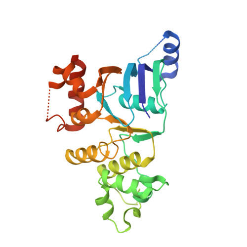
Structure and dynamics of NBD1 from CFTR characterized using crystallography and hydrogen/deuterium exchange mass spectrometry.
Lewis, H.A., Wang, C., Zhao, X., Hamuro, Y., Conners, K., Kearins, M.C., Lu, F., Sauder, J.M., Molnar, K.S., Coales, S.J., Maloney, P.C., Guggino, W.B., Wetmore, D.R., Weber, P.C., Hunt, J.F.(2010) J Mol Biology 396: 406-430
- PubMed: 19944699 Search on PubMed
- DOI: https://doi.org/10.1016/j.jmb.2009.11.051
- Primary Citation Related Structures:
2BBO, 2BBS, 2BBT - PubMed Abstract:
The DeltaF508 mutation in nucleotide-binding domain 1 (NBD1) of the cystic fibrosis transmembrane conductance regulator (CFTR) is the predominant cause of cystic fibrosis. Previous biophysical studies on human F508 and DeltaF508 domains showed only local structural changes restricted to residues 509-511 and only minor differences in folding rate and stability. These results were remarkable because DeltaF508 was widely assumed to perturb domain folding based on the fact that it prevents trafficking of CFTR out of the endoplasmic reticulum. However, the previously reported crystal structures did not come from matched F508 and DeltaF508 constructs, and the DeltaF508 structure contained additional mutations that were required to obtain sufficient protein solubility. In this article, we present additional biophysical studies of NBD1 designed to address these ambiguities. Mass spectral measurements of backbone amide (1)H/(2)H exchange rates in matched F508 and DeltaF508 constructs reveal that DeltaF508 increases backbone dynamics at residues 509-511 and the adjacent protein segments but not elsewhere in NBD1. These measurements also confirm a high level of flexibility in the protein segments exhibiting variable conformations in the crystal structures. We additionally present crystal structures of a broader set of human NBD1 constructs, including one harboring the native F508 residue and others harboring the DeltaF508 mutation in the presence of fewer and different solubilizing mutations. The only consistent conformational difference is observed at residues 509-511. The side chain of residue V510 in this loop is mostly buried in all non-DeltaF508 structures but completely solvent exposed in all DeltaF508 structures. These results reinforce the importance of the perturbation DeltaF508 causes in the surface topography of NBD1 in a region likely to mediate contact with the transmembrane domains of CFTR. However, they also suggest that increased exposure of the 509-511 loop and increased dynamics in its vicinity could promote aggregation in vitro and aberrant intermolecular interactions that impede trafficking in vivo.
- SGX Pharmaceuticals, San Diego, CA 92121, USA.
Organizational Affiliation: