2B7S | pdb_00002b7s

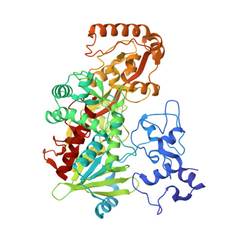
R381K mutant of flavocytochrome c3


Experimental Data Snapshot

  • Method: X-RAY DIFFRACTION
  • Resolution: 2.12 Å
  • R-Value Free: 
    0.197 (Depositor), 0.197 (DCC) 
  • R-Value Work: 
    0.144 (Depositor), 0.145 (DCC) 
  • R-Value Observed: 
    0.147 (Depositor) 

Starting Model: experimental
View more details

wwPDB Validation 3D Report Full Report

Validation slider image for 2B7S

Ligand Structure Quality Assessment 


This is version 4.0 of the entry. See complete history. 

Literature

A Proton Delivery Pathway in the Soluble Fumarate Reductase from Shewanella frigidimarina.

Pankhurst, K.L., Mowat, C.G., Rothery, E.L., Hudson, J.M., Jones, A.K., Miles, C.S., Walkinshaw, M.D., Armstrong, F.A., Reid, G.A., Chapman, S.K.

(2006) J Biol Chem 281: 20589-20597

  • DOI: https://doi.org/10.1074/jbc.M603077200
  • Primary Citation Related Structures: 
    2B7R, 2B7S

  • PubMed Abstract: 

    The mechanism for fumarate reduction by the soluble fumarate reductase from Shewanella frigidimarina involves hydride transfer from FAD and proton transfer from the active-site acid, Arg-402. It has been proposed that Arg-402 forms part of a proton transfer pathway that also involves Glu-378 and Arg-381 but, unusually, does not involve any bound water molecules. To gain further insight into the importance of this proton pathway we have perturbed it by substituting Arg-381 by lysine and methionine and Glu-378 by aspartate. Although all the mutant enzymes retain measurable activities, there are orders-of-magnitude decreases in their k(cat) values compared with the wild-type enzyme. Solvent kinetic isotope effects show that proton transfer is rate-limiting in the wild-type and mutant enzymes. Proton inventories indicate that the proton pathway involves multiple exchangeable groups. Fast scan protein-film voltammetric studies on wild-type and R381K enzymes show that the proton transfer pathway delivers one proton per catalytic cycle and is not required for transporting the other proton, which transfers as a hydride from the reduced, protonated FAD. The crystal structures of E378D and R381M mutant enzymes have been determined to 1.7 and 2.1 A resolution, respectively. They allow an examination of the structural changes that disturb proton transport. Taken together, the results indicate that Arg-381, Glu-378, and Arg-402 form a proton pathway that is completely conserved throughout the fumarate reductase/succinate dehydrogenase family of enzymes.


  • Organizational Affiliation: 
    • Edinburgh and St. Andrews Research School of Chemistry, University of Edinburgh, West Mains Road, Edinburgh EH9 3JJ, UK.

Macromolecule Content 

  • Total Structure Weight: 64.01 kDa 
  • Atom Count: 5,192 
  • Modeled Residue Count: 568 
  • Deposited Residue Count: 571 
  • Unique protein chains: 1

Macromolecules

Find similar proteins by:|  3D Structure
Entity ID: 1
MoleculeChains  Sequence LengthOrganismDetailsImage
Fumarate reductase flavoprotein subunit571Shewanella frigidimarinaMutation(s): 1 
Gene Names: FCC
EC: 1.3.99.1 (PDB Primary Data), 1.3.2.4 (UniProt)
UniProt
Find proteins for P0C278 (Shewanella frigidimarina)
Explore P0C278 
Go to UniProtKB:  P0C278
Entity Groups
Sequence Clusters30% Identity50% Identity70% Identity90% Identity95% Identity100% Identity
UniProt GroupP0C278
Sequence Annotations
Expand
Reference Sequence

Small Molecules

Ligands 4 Unique
IDChains Name / Formula / InChI Key2D Diagram3D Interactions
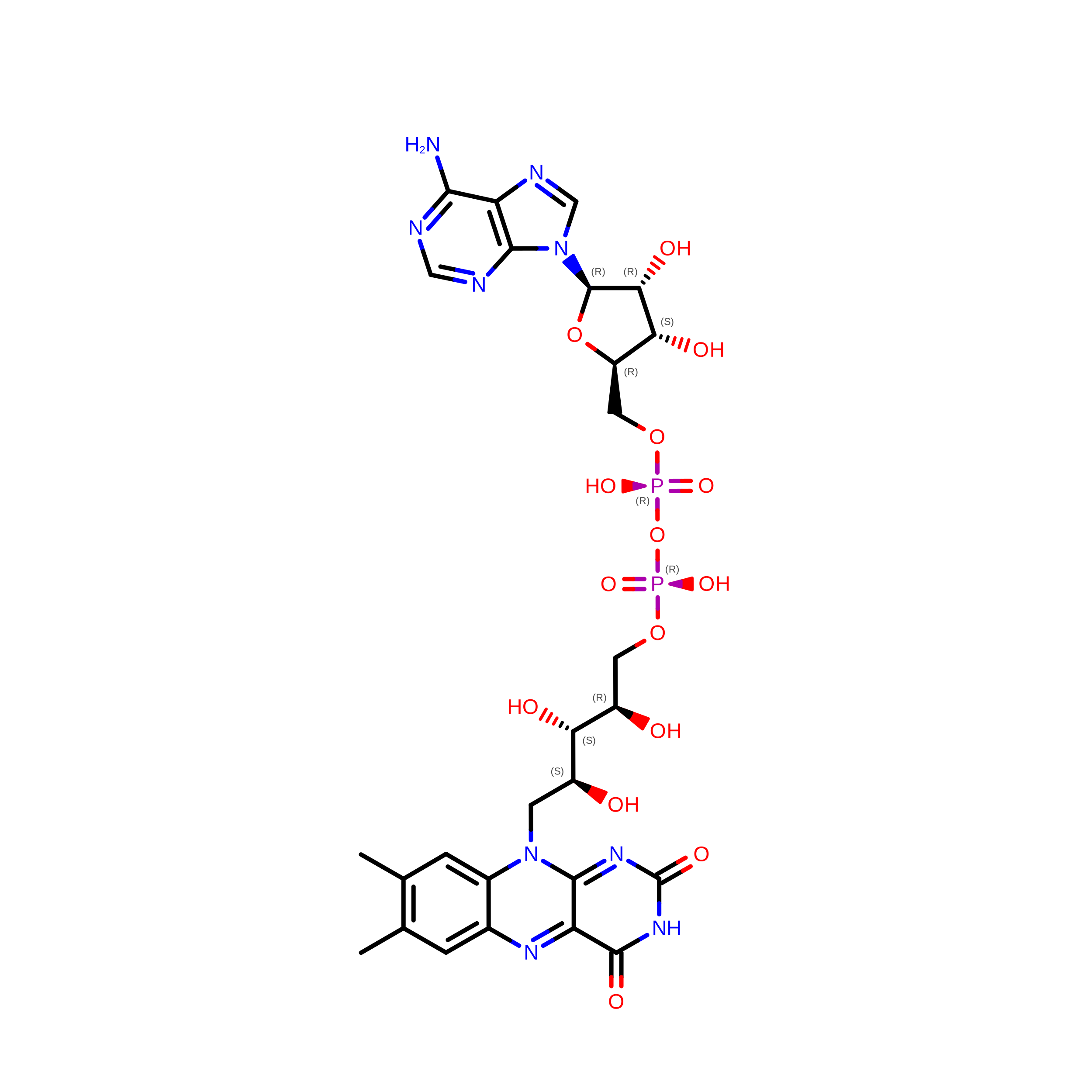
FAD

Query on FAD


Download:Ideal Coordinates CCD File
G [auth A]FLAVIN-ADENINE DINUCLEOTIDE
C27 H33 N9 O15 P2
VWWQXMAJTJZDQX-UYBVJOGSSA-N
HEC

Query on HEC


Download:Ideal Coordinates CCD File
C [auth A],
D [auth A],
E [auth A],
F [auth A]
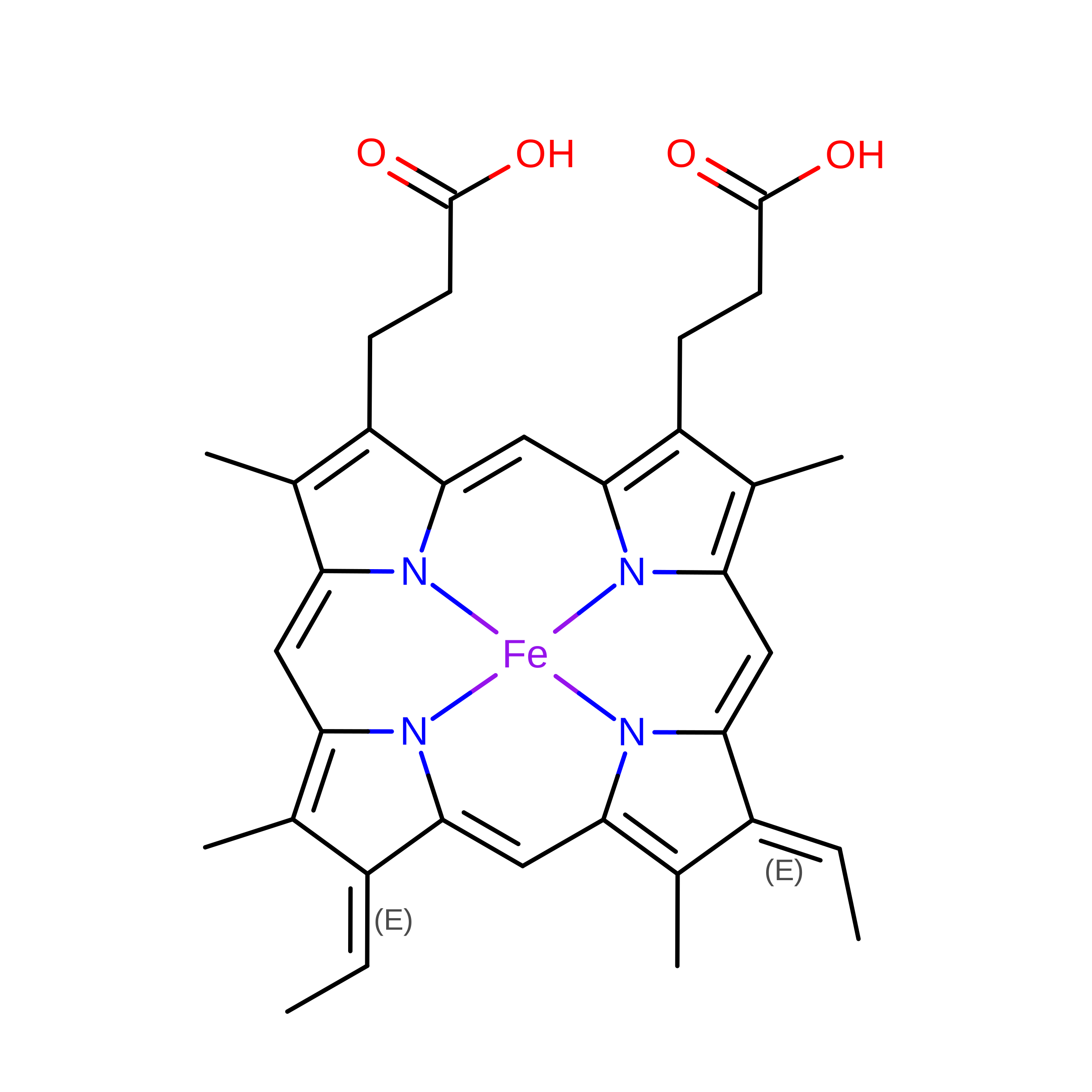
HEME C
C34 H36 Fe N4 O4
YZTICFPLOXKSQO-RGGAHWMASA-L
FUM

Query on FUM


Download:Ideal Coordinates CCD File
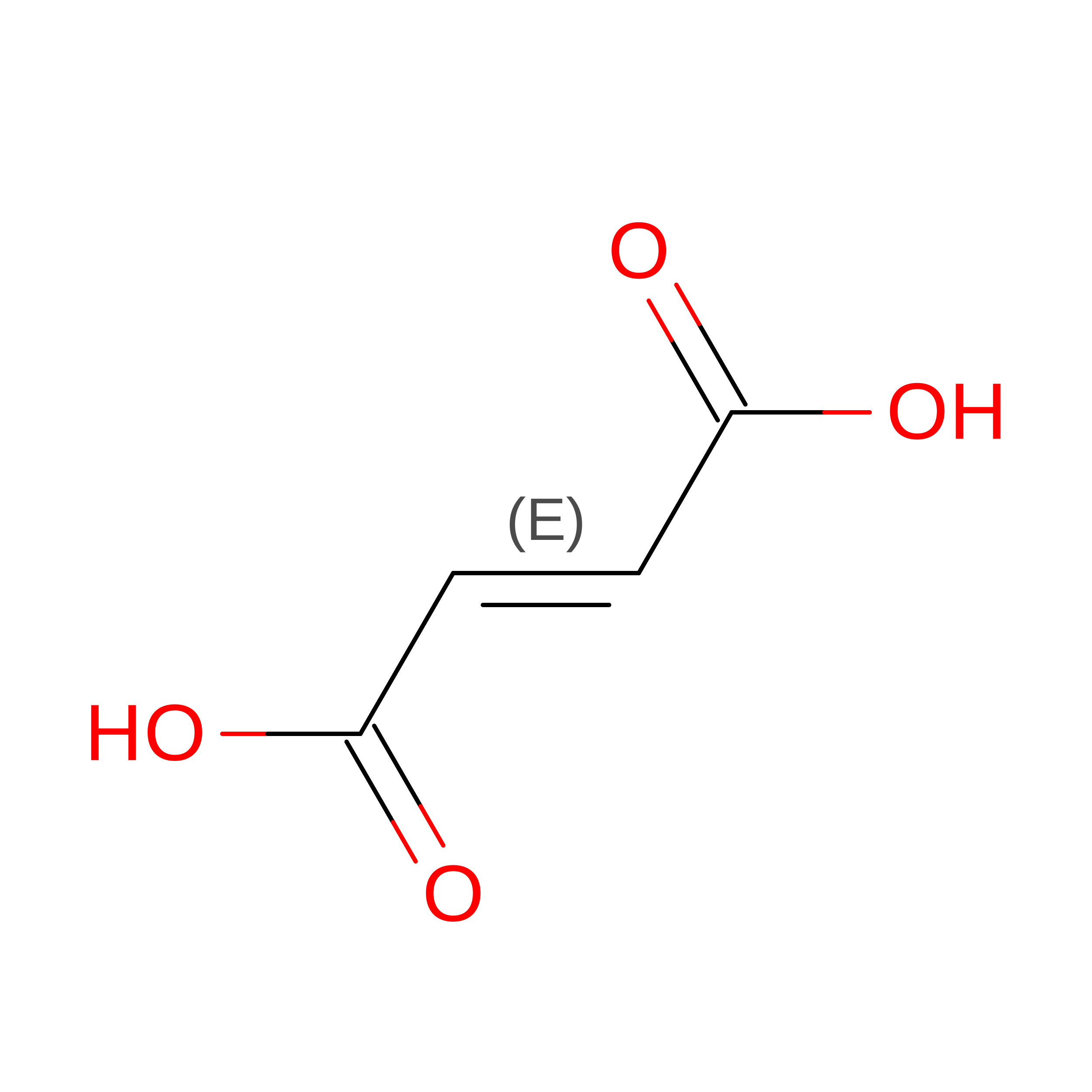
H [auth A]FUMARIC ACID
C4 H4 O4
VZCYOOQTPOCHFL-OWOJBTEDSA-N
NA

Query on NA


Download:Ideal Coordinates CCD File
B [auth A]SODIUM ION
Na
FKNQFGJONOIPTF-UHFFFAOYSA-N

Experimental Data & Validation

Experimental Data

  • Method: X-RAY DIFFRACTION
  • Resolution: 2.12 Å
  • R-Value Free:  0.197 (Depositor), 0.197 (DCC) 
  • R-Value Work:  0.144 (Depositor), 0.145 (DCC) 
  • R-Value Observed: 0.147 (Depositor) 
Space Group: P 1 21 1
Unit Cell:
Length ( Å )Angle ( ˚ )
a = 45.228α = 90
b = 91.362β = 91.26
c = 78.193γ = 90
Software Package:
Software NamePurpose
REFMACrefinement
DENZOdata reduction
SCALEPACKdata scaling
AMoREphasing

Structure Validation

View Full Validation Report


Ligand Structure Quality Assessment 


Entry History 

Deposition Data

Revision History  (Full details and data files)

  • Version 1.0: 2006-05-23
    Type: Initial release
  • Version 1.1: 2008-05-01
    Changes: Version format compliance
  • Version 1.2: 2011-07-13
    Changes: Version format compliance
  • Version 1.3: 2021-11-10
    Changes: Database references, Derived calculations
  • Version 1.4: 2023-10-25
    Changes: Data collection, Refinement description
  • Version 2.0: 2023-11-15
    Changes: Atomic model, Data collection
  • Version 2.1: 2024-11-20
    Changes: Structure summary
  • Version 3.0: 2025-12-24
    Changes: Advisory, Atomic model, Data collection, Derived calculations, Non-polymer description, Structure summary
  • Version 4.0: 2026-08-12
    Type: Remediation
    Reason: Metalloprotein remediation
    Changes: Advisory, Data collection, Derived calculations, Non-polymer description, Structure summary