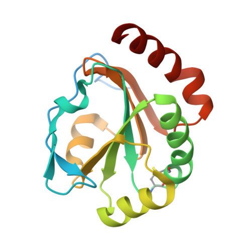
Crystal structures of E. coli CcmG and its mutants reveal key roles of the N-terminal beta-sheet and the fingerprint region
Ouyang, N., Gao, Y.G., Hu, H.Y., Xia, Z.X.(2006) Proteins 65: 1021-1031
- PubMed: 17019698 Search on PubMed
- DOI: https://doi.org/10.1002/prot.21184
- Primary Citation Related Structures:
2B1K, 2B1L, 2G0F - PubMed Abstract:
CcmG, also designated DsbE, functions as a periplasmic protein thiol:disulfide oxidoreductase and is required for cytochrome c maturation. Here we report the crystal structures of Escherichia coli CcmG and its two mutants, P144A and the N-terminal fifty seven-residue deletion mutant, and two additional deletion mutants were studied by circular dichroism. Structural comparison of E. coli CcmG with its deletion mutants reveals that the N-terminal beta-sheet is essential for maintaining the folding topology and consequently maintaining the active-site structure of CcmG. Pro144 and Glu145 are key residues of the fingerprint region of CcmG. Pro144 is in cis-configuration, and it makes van der Waals interactions with the active-site disulfide Cys80-Cys83 and forms a C--H...O hydrogen bond with Thr82, helping stabilize the active-site structure. Glu145 forms a salt-bridge and hydrogen-bond network with other residues of the fingerprint region and with Arg158, further stabilizing the active-site structure. The cis-configuration of Pro144 makes the backbone nitrogen and oxygen of Ala143 exposed to solvent, favorable for interacting with binding partners. The key role of cis-Pro144 is verified by the P144A mutant, which contains trans-Ala144 and displays redox property changes. Structural comparison of E. coli CcmG with the recently reported structure of CcmG in complex with the N-terminal domain of DsbD reveals that Tyr141 undergoes conformational changes upon binding DsbD. A cis-proline located at the N-terminus of the first beta-strand of the betabetaalpha motif of the thioredoxin-like domain is a conserved structural feature of the thioredoxin superfamily.
- State Key Laboratory of Bio-organic and Natural Products Chemistry, Shanghai Institute of Organic Chemistry, Chinese Academy of Sciences, Shanghai 200032, China.
Organizational Affiliation: