2B1C

5'-D(*GP*CP*GP*TP*GP*GP*GP*AP*CP*C)-3' Zif268 binding site


Experimental Data Snapshot

  • Method: X-RAY DIFFRACTION
  • Resolution: 2.20 Å
  • R-Value Free: 0.286 
  • R-Value Work: 0.234 
  • R-Value Observed: 0.234 

wwPDB Validation   3D Report Full Report


This is version 1.4 of the entry. See complete history


Literature

Signatures of protein-DNA recognition in free DNA binding sites.

Locasale, J.W.Napoli, A.A.Chen, S.Berman, H.M.Lawson, C.L.

(2009) J Mol Biol 386: 1054-1065

  • DOI: https://doi.org/10.1016/j.jmb.2009.01.007
  • Primary Citation of Related Structures:  
    1HQ7, 2B1B, 2B1C, 2B1D

  • PubMed Abstract: 

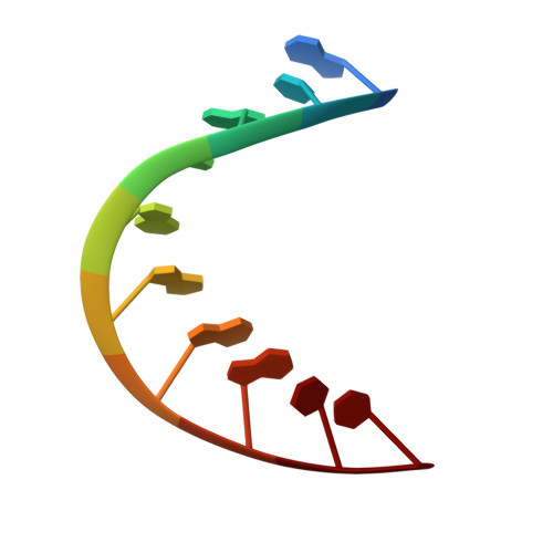
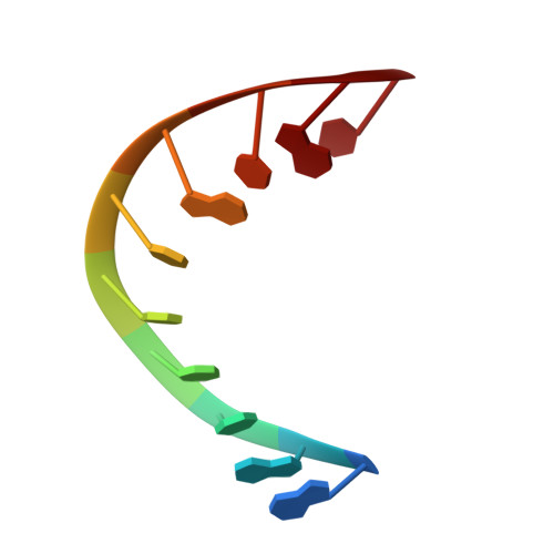
    One obstacle to achieving complete understanding of the principles underlying sequence-dependent recognition of DNA is the paucity of structural data for DNA recognition sequences in their free (unbound) state. Here, we carried out crystallization screening of 50 DNA duplexes containing cognate protein binding sites and obtained new crystal structures of free DNA binding sites for three distinct modes of DNA recognition: anti-parallel beta strands (MetR), helix-turn-helix motif + hinge helices (PurR), and zinc fingers (Zif268). Structural changes between free and protein-bound DNA are manifested differently in each case. The new DNA structures reveal that distinctive sequence-dependent DNA geometry dominates recognition by MetR, protein-induced bending of DNA dictates recognition by PurR, and deformability of DNA along the A-B continuum is important in recognition by Zif268. Together, our findings show that crystal structures of free DNA binding sites provide new information about the nature of protein-DNA interactions and thus lend insights towards a structural code for DNA recognition.


  • Organizational Affiliation

    Department of Chemistry and Chemical Biology, Rutgers, State University of New Jersey, 610 Taylor Road, Piscataway, NJ 08854, USA. cathy.lawson@rutgers.edu


Macromolecules

Find similar nucleic acids by:  Sequence   |   3D Structure  

Entity ID: 1
MoleculeChains LengthOrganismImage
5'-D(*GP*CP*GP*TP*GP*GP*GP*AP*CP*C)-3'10N/A
Sequence Annotations
Expand
  • Reference Sequence

Find similar nucleic acids by:  Sequence   |   3D Structure  

Entity ID: 2
MoleculeChains LengthOrganismImage
5'-D(*GP*GP*TP*CP*CP*CP*AP*CP*GP*C)-3'10N/A
Sequence Annotations
Expand
  • Reference Sequence
Small Molecules
Ligands 1 Unique
IDChains Name / Formula / InChI Key2D Diagram3D Interactions
MG
Query on MG

Download Ideal Coordinates CCD File 
C [auth A]MAGNESIUM ION
Mg
JLVVSXFLKOJNIY-UHFFFAOYSA-N
Experimental Data & Validation

Experimental Data

  • Method: X-RAY DIFFRACTION
  • Resolution: 2.20 Å
  • R-Value Free: 0.286 
  • R-Value Work: 0.234 
  • R-Value Observed: 0.234 
  • Space Group: P 61 2 2
Unit Cell:
Length ( Å )Angle ( ˚ )
a = 38.482α = 90
b = 38.482β = 90
c = 78.225γ = 120
Software Package:
Software NamePurpose
DENZOdata reduction
SCALEPACKdata scaling
AMoREphasing
CNSrefinement
PDB_EXTRACTdata extraction
CBASSdata collection

Structure Validation

View Full Validation Report


Entry History 

Deposition Data

Revision History  (Full details and data files)

  • Version 1.0: 2005-09-27
    Type: Initial release
  • Version 1.1: 2008-05-01
    Changes: Version format compliance
  • Version 1.2: 2011-07-13
    Changes: Version format compliance
  • Version 1.3: 2017-10-11
    Changes: Refinement description
  • Version 1.4: 2023-08-23
    Changes: Data collection, Database references, Derived calculations, Refinement description