2B0U

The Structure of the Follistatin:Activin Complex


Experimental Data Snapshot

  • Method: X-RAY DIFFRACTION
  • Resolution: 2.80 Å
  • R-Value Free: 0.297 
  • R-Value Work: 0.265 
  • R-Value Observed: 0.301 

wwPDB Validation   3D Report Full Report


This is version 1.4 of the entry. See complete history


Literature

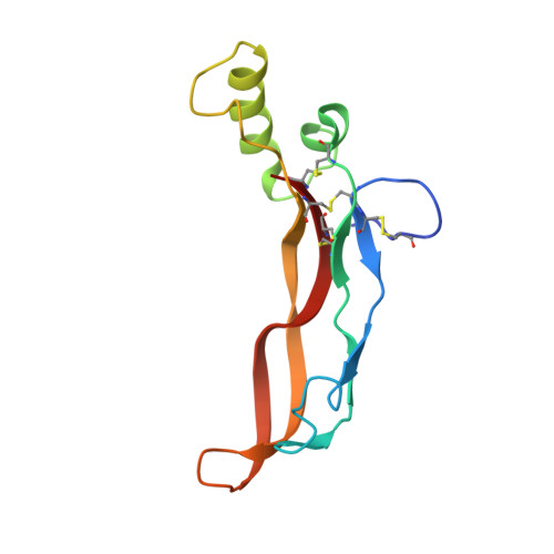
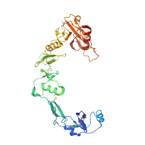
The Structure of the Follistatin:Activin Complex Reveals Antagonism of Both Type I and Type II Receptor Binding.

Thompson, T.B.Lerch, T.F.Cook, R.W.Woodruff, T.K.Jardetzky, T.S.

(2005) Dev Cell 9: 535-543

  • DOI: https://doi.org/10.1016/j.devcel.2005.09.008
  • Primary Citation of Related Structures:  
    2B0U

  • PubMed Abstract: 

    TGF-beta ligands stimulate diverse cellular differentiation and growth responses by signaling through type I and II receptors. Ligand antagonists, such as follistatin, block signaling and are essential regulators of physiological responses. Here we report the structure of activin A, a TGF-beta ligand, bound to the high-affinity antagonist follistatin. Two follistatin molecules encircle activin, neutralizing the ligand by burying one-third of its residues and its receptor binding sites. Previous studies have suggested that type I receptor binding would not be blocked by follistatin, but the crystal structure reveals that the follistatin N-terminal domain has an unexpected fold that mimics a universal type I receptor motif and occupies this receptor binding site. The formation of follistatin:BMP:type I receptor complexes can be explained by the stoichiometric and geometric arrangement of the activin:follistatin complex. The mode of ligand binding by follistatin has important implications for its ability to neutralize homo- and heterodimeric ligands of this growth factor family.


  • Organizational Affiliation

    Department of Biochemistry, Northwestern University, Evanston, Illinois 60208, USA.


Macromolecules
Find similar proteins by:  (by identity cutoff)  |  3D Structure
Entity ID: 1
MoleculeChains Sequence LengthOrganismDetailsImage
Inhibin beta A chain
A, B
116Homo sapiensMutation(s): 0 
Gene Names: INHBA
UniProt & NIH Common Fund Data Resources
Find proteins for P08476 (Homo sapiens)
Explore P08476 
Go to UniProtKB:  P08476
PHAROS:  P08476
GTEx:  ENSG00000122641 
Entity Groups  
Sequence Clusters30% Identity50% Identity70% Identity90% Identity95% Identity100% Identity
UniProt GroupP08476
Sequence Annotations
Expand
  • Reference Sequence
Find similar proteins by:  (by identity cutoff)  |  3D Structure
Entity ID: 2
MoleculeChains Sequence LengthOrganismDetailsImage
Follistatin
C, D
288Homo sapiensMutation(s): 0 
Gene Names: FST
UniProt & NIH Common Fund Data Resources
Find proteins for P19883 (Homo sapiens)
Explore P19883 
Go to UniProtKB:  P19883
PHAROS:  P19883
GTEx:  ENSG00000134363 
Entity Groups  
Sequence Clusters30% Identity50% Identity70% Identity90% Identity95% Identity100% Identity
UniProt GroupP19883
Sequence Annotations
Expand
  • Reference Sequence
Small Molecules
Ligands 3 Unique
IDChains Name / Formula / InChI Key2D Diagram3D Interactions
IR3
Query on IR3

Download Ideal Coordinates CCD File 
E [auth A],
I [auth B],
K [auth C],
L [auth C],
N [auth D]
IRIDIUM (III) ION
Ir
MILUBEOXRNEUHS-UHFFFAOYSA-N
MPD
Query on MPD

Download Ideal Coordinates CCD File 
J [auth B](4S)-2-METHYL-2,4-PENTANEDIOL
C6 H14 O2
SVTBMSDMJJWYQN-YFKPBYRVSA-N
MLI
Query on MLI

Download Ideal Coordinates CCD File 
F [auth A],
G [auth A],
H [auth A],
M [auth C]
MALONATE ION
C3 H2 O4
OFOBLEOULBTSOW-UHFFFAOYSA-L
Experimental Data & Validation

Experimental Data

  • Method: X-RAY DIFFRACTION
  • Resolution: 2.80 Å
  • R-Value Free: 0.297 
  • R-Value Work: 0.265 
  • R-Value Observed: 0.301 
  • Space Group: P 21 21 2
Unit Cell:
Length ( Å )Angle ( ˚ )
a = 109.25α = 90
b = 120.82β = 90
c = 70.57γ = 90
Software Package:
Software NamePurpose
SCALAdata scaling
SHARPphasing
CNSrefinement
PDB_EXTRACTdata extraction
MAR345data collection
CCP4data scaling

Structure Validation

View Full Validation Report


Entry History 

Deposition Data

Revision History  (Full details and data files)

  • Version 1.0: 2005-10-11
    Type: Initial release
  • Version 1.1: 2008-05-01
    Changes: Version format compliance
  • Version 1.2: 2011-07-13
    Changes: Version format compliance
  • Version 1.3: 2011-07-27
    Changes: Version format compliance
  • Version 1.4: 2017-10-11
    Changes: Refinement description