2AW2 | pdb_00002aw2

Crystal structure of the human BTLA-HVEM complex


Experimental Data Snapshot

  • Method: X-RAY DIFFRACTION
  • Resolution: 2.80 Å
  • R-Value Free: 
    0.278 (Depositor), 0.281 (DCC) 
  • R-Value Work: 
    0.231 (Depositor) 
  • R-Value Observed: 
    0.236 (Depositor) 

Starting Models: experimental
View more details

wwPDB Validation 3D Report Full Report

Validation slider image for 2AW2

This is version 2.2 of the entry. See complete history

Literature

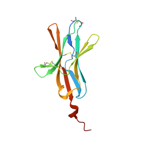
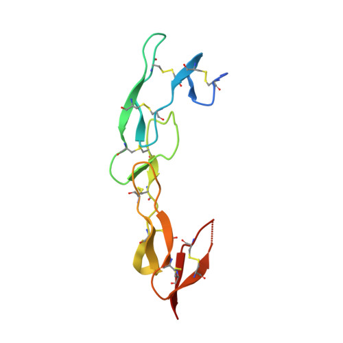
Attenuating Lymphocyte Activity: the crystal structure of the BTLA-HVEM complex

Compaan, D.M.Gonzalez, L.C.Tom, I.Loyet, K.M.Eaton, D.Hymowitz, S.G.

(2005) J Biological Chem 280: 39553-39561

  • DOI: https://doi.org/10.1074/jbc.M507629200
  • Primary Citation Related Structures: 
    2AW2

  • PubMed Abstract: 

    Five CD28-like proteins exert positive or negative effects on immune cells. Only four of these five receptors interact with members of the B7 family. The exception is BTLA (B and T lymphocyte attenuator), which instead interacts with the tumor necrosis factor receptor superfamily member HVEM (herpes virus entry mediator). To better understand this interaction, we determined the 2.8-A crystal structure of the BTLA-HVEM complex. This structure shows that BTLA binds the N-terminal cysteine-rich domain of HVEM and employs a unique binding surface compared with other CD28-like receptors. Moreover, the structure shows that BTLA recognizes the same surface on HVEM as gD (herpes virus glycoprotein D) and utilizes a similar binding motif. Light scattering analysis demonstrates that the extracellular domain of BTLA is monomeric and that BTLA and HVEM form a 1:1 complex. Alanine-scanning mutagenesis of HVEM was used to further define critical binding residues. Finally, BTLA adopts an immunoglobulin I-set fold. Despite structural similarities to other CD28-like members, BTLA represents a unique co-receptor.


  • Organizational Affiliation
    • Department of Protein Engineering, Genentech, Inc., S. San Francisco, California 94080, USA.

Macromolecule Content 

  • Total Structure Weight: 51.96 kDa 
  • Atom Count: 3,259 
  • Modeled Residue Count: 417 
  • Deposited Residue Count: 456 
  • Unique protein chains: 2

Macromolecules

Find similar proteins by:|  3D Structure
Entity ID: 1
MoleculeChains  Sequence LengthOrganismDetailsImage
B and T lymphocyte attenuatorA,
C [auth X]
120Homo sapiensMutation(s): 0 
Gene Names: BTLA
UniProt & NIH Common Fund Data Resources
Find proteins for Q7Z6A9 (Homo sapiens)
Explore Q7Z6A9 
Go to UniProtKB:  Q7Z6A9
PHAROS:  Q7Z6A9
GTEx:  ENSG00000186265 
Entity Groups
Sequence Clusters30% Identity50% Identity70% Identity90% Identity95% Identity100% Identity
UniProt GroupQ7Z6A9
Sequence Annotations
Expand
Reference Sequence
Find similar proteins by:|  3D Structure
Entity ID: 2
MoleculeChains  Sequence LengthOrganismDetailsImage
Tumor necrosis factor receptor superfamily member 14B,
D [auth Y]
108Homo sapiensMutation(s): 0 
Gene Names: TNFRSF14HVEAHVEM
UniProt & NIH Common Fund Data Resources
Find proteins for Q92956 (Homo sapiens)
Explore Q92956 
Go to UniProtKB:  Q92956
PHAROS:  Q92956
GTEx:  ENSG00000157873 
Entity Groups
Sequence Clusters30% Identity50% Identity70% Identity90% Identity95% Identity100% Identity
UniProt GroupQ92956
Glycosylation
Glycosylation Sites: 1Go to GlyGen: Q92956-1
Sequence Annotations
Expand
Reference Sequence

Oligosaccharides

Help  
Entity ID: 3
MoleculeChains Length2D Diagram GlycosylationD Interactions
2-acetamido-2-deoxy-beta-D-glucopyranose-(1-4)-2-acetamido-2-deoxy-beta-D-glucopyranoseE [auth C]2N-Glycosylation
Glycosylation Resources
GlyTouCan: G42666HT
GlyCosmos: G42666HT
GlyGen: G42666HT
Entity ID: 4
MoleculeChains Length2D Diagram GlycosylationD Interactions
beta-L-fucopyranose-(1-6)-2-acetamido-2-deoxy-beta-D-glucopyranoseF [auth D]2N-Glycosylation
Glycosylation Resources
GlyTouCan: G61843VN
GlyCosmos: G61843VN
GlyGen: G61843VN

Small Molecules

Ligands 1 Unique
IDChains Name / Formula / InChI Key2D Diagram3D Interactions
NI

Query on NI


Download:Ideal Coordinates CCD File
G [auth A]NICKEL (II) ION
Ni
VEQPNABPJHWNSG-UHFFFAOYSA-N

Experimental Data & Validation

Experimental Data

  • Method: X-RAY DIFFRACTION
  • Resolution: 2.80 Å
  • R-Value Free:  0.278 (Depositor), 0.281 (DCC) 
  • R-Value Work:  0.231 (Depositor) 
  • R-Value Observed: 0.236 (Depositor) 
Space Group: C 2 2 21
Unit Cell:
Length ( Å )Angle ( ˚ )
a = 49.294α = 90
b = 167.116β = 90
c = 148.68γ = 90
Software Package:
Software NamePurpose
REFMACrefinement
HKL-2000data reduction
SCALEPACKdata scaling
PHASERphasing

Structure Validation

View Full Validation Report


Entry History 

Deposition Data

Revision History  (Full details and data files)

  • Version 1.0: 2005-09-27
    Type: Initial release
  • Version 1.1: 2008-05-01
    Changes: Version format compliance
  • Version 1.2: 2011-07-13
    Changes: Advisory, Refinement description, Version format compliance
  • Version 2.0: 2020-07-29
    Type: Remediation
    Reason: Carbohydrate remediation
    Changes: Atomic model, Data collection, Database references, Derived calculations, Structure summary
  • Version 2.1: 2023-08-23
    Changes: Data collection, Database references, Refinement description, Structure summary
  • Version 2.2: 2024-10-30
    Changes: Structure summary