2AV1

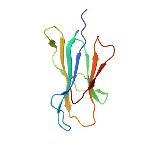
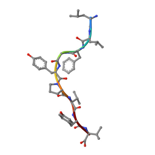
Crystal structure of HTLV-1 TAX peptide Bound to Human Class I MHC HLA-A2 with the E63Q and K66A mutations in the heavy chain.


Experimental Data Snapshot

  • Method: X-RAY DIFFRACTION
  • Resolution: 1.95 Å
  • R-Value Free: 0.231 
  • R-Value Work: 0.174 
  • R-Value Observed: 0.177 

Starting Model: experimental
View more details

wwPDB Validation   3D Report Full Report


This is version 1.6 of the entry. See complete history


Literature

Unraveling a Hotspot for TCR Recognition on HLA-A2: Evidence Against the Existence of Peptide-independent TCR Binding Determinants.

Gagnon, S.J.Borbulevych, O.Y.Davis-Harrison, R.L.Baxter, T.K.Clemens, J.R.Armstrong, K.M.Turner, R.V.Damirjian, M.Biddison, W.E.Baker, B.M.

(2005) J Mol Biol 353: 556-573

  • DOI: https://doi.org/10.1016/j.jmb.2005.08.024
  • Primary Citation of Related Structures:  
    2AV1, 2AV7

  • PubMed Abstract: 

    T cell receptor (TCR) recognition of peptide takes place in the context of the major histocompatibility complex (MHC) molecule, which accounts for approximately two-thirds of the peptide/MHC buried surface. Using the class I MHC HLA-A2 and a large panel of mutants, we have previously shown that surface mutations that disrupt TCR recognition vary with the identity of the peptide. The single exception is Lys66 on the HLA-A2 alpha1 helix, which when mutated to alanine disrupts recognition for 93% of over 250 different T cell clones or lines, independent of which peptide is bound. Thus, Lys66 could serve as a peptide-independent TCR binding determinant. Here, we have examined the role of Lys66 in TCR recognition of HLA-A2 in detail. The structure of a peptide/HLA-A2 molecule with the K66A mutation indicates that although the mutation induces no major structural changes, it results in the exposure of a negatively charged glutamate (Glu63) underneath Lys66. Concurrent replacement of Glu63 with glutamine restores TCR binding and function for T cells specific for five different peptides presented by HLA-A2. Thus, the positive charge on Lys66 does not serve to guide all TCRs onto the HLA-A2 molecule in a manner required for productive signaling. Furthermore, electrostatic calculations indicate that Lys66 does not contribute to the stability of two TCR-peptide/HLA-A2 complexes. Our findings are consistent with the notion that each TCR arrives at a unique solution of how to bind a peptide/MHC, most strongly influenced by the chemical and structural features of the bound peptide. This would not rule out an intrinsic affinity of TCRs for MHC molecules achieved through multiple weak interactions, but for HLA-A2 the collective mutational data place limits on the role of any single MHC amino acid side-chain in driving TCR binding in a peptide-independent fashion.


  • Organizational Affiliation

    Molecular Immunology Section, Neuroimmunology Branch, National Institute of Neurological Diseases and Stroke, National Institutes of Health, Bethesda, MD 20892, USA.


Macromolecules
Find similar proteins by:  (by identity cutoff)  |  3D Structure
Entity ID: 1
MoleculeChains Sequence LengthOrganismDetailsImage
HLA class I histocompatibility antigen, A-2 alpha chain
A, D
275Homo sapiensMutation(s): 2 
Gene Names: HLA-AHLAA
UniProt & NIH Common Fund Data Resources
Find proteins for P04439 (Homo sapiens)
Explore P04439 
Go to UniProtKB:  P04439
PHAROS:  P04439
GTEx:  ENSG00000206503 
Entity Groups  
Sequence Clusters30% Identity50% Identity70% Identity90% Identity95% Identity100% Identity
UniProt GroupP04439
Sequence Annotations
Expand
  • Reference Sequence
Find similar proteins by:  (by identity cutoff)  |  3D Structure
Entity ID: 2
MoleculeChains Sequence LengthOrganismDetailsImage
Beta-2-microglobulin
B, E
100Homo sapiensMutation(s): 0 
Gene Names: B2M
UniProt & NIH Common Fund Data Resources
Find proteins for P61769 (Homo sapiens)
Explore P61769 
Go to UniProtKB:  P61769
PHAROS:  P61769
GTEx:  ENSG00000166710 
Entity Groups  
Sequence Clusters30% Identity50% Identity70% Identity90% Identity95% Identity100% Identity
UniProt GroupP61769
Sequence Annotations
Expand
  • Reference Sequence

Find similar proteins by:  Sequence   |   3D Structure  

Entity ID: 3
MoleculeChains Sequence LengthOrganismDetailsImage
Trans-activating transcriptional regulatory peptide
C, F
9Human T-cell lymphotrophic virus type 1 (Caribbean isolate)Mutation(s): 0 
UniProt
Find proteins for P14079 (Human T-cell leukemia virus 1 (isolate Caribbea HS-35 subtype A))
Explore P14079 
Go to UniProtKB:  P14079
Entity Groups  
UniProt GroupP14079
Sequence Annotations
Expand
  • Reference Sequence
Small Molecules
Ligands 2 Unique
IDChains Name / Formula / InChI Key2D Diagram3D Interactions
GOL
Query on GOL

Download Ideal Coordinates CCD File 
AA [auth D]
BA [auth E]
CA [auth E]
DA [auth E]
G [auth A]
AA [auth D],
BA [auth E],
CA [auth E],
DA [auth E],
G [auth A],
H [auth A],
I [auth A],
J [auth A],
K [auth A],
L [auth A],
N [auth B],
O [auth B],
P [auth B],
Q [auth B],
R [auth B],
S [auth B],
T [auth C],
U [auth D],
V [auth D],
W [auth D],
X [auth D],
Y [auth D],
Z [auth D]
GLYCEROL
C3 H8 O3
PEDCQBHIVMGVHV-UHFFFAOYSA-N
SCN
Query on SCN

Download Ideal Coordinates CCD File 
M [auth B]THIOCYANATE ION
C N S
ZMZDMBWJUHKJPS-UHFFFAOYSA-M
Experimental Data & Validation

Experimental Data

  • Method: X-RAY DIFFRACTION
  • Resolution: 1.95 Å
  • R-Value Free: 0.231 
  • R-Value Work: 0.174 
  • R-Value Observed: 0.177 
  • Space Group: P 1
Unit Cell:
Length ( Å )Angle ( ˚ )
a = 50.328α = 82.1
b = 62.536β = 76.39
c = 74.747γ = 78.1
Software Package:
Software NamePurpose
REFMACrefinement
HKL-2000data reduction
HKL-2000data scaling
MOLREPphasing

Structure Validation

View Full Validation Report


Entry History 

Deposition Data

Revision History  (Full details and data files)

  • Version 1.0: 2005-10-18
    Type: Initial release
  • Version 1.1: 2008-05-01
    Changes: Version format compliance
  • Version 1.2: 2011-07-13
    Changes: Advisory, Version format compliance
  • Version 1.3: 2012-12-12
    Changes: Other
  • Version 1.4: 2013-03-13
    Changes: Other
  • Version 1.5: 2021-10-20
    Changes: Database references, Derived calculations
  • Version 1.6: 2023-08-23
    Changes: Data collection, Refinement description