2AU3

Crystal Structure of the Aquifex aeolicus primase (Zinc Binding and RNA Polymerase Domains)


Experimental Data Snapshot

  • Method: X-RAY DIFFRACTION
  • Resolution: 2.00 Å
  • R-Value Free: 0.238 
  • R-Value Work: 0.203 
  • R-Value Observed: 0.205 

wwPDB Validation   3D Report Full Report


This is version 1.3 of the entry. See complete history


Literature

Crosstalk between primase subunits can act to regulate primer synthesis in trans.

Corn, J.E.Pease, P.J.Hura, G.L.Berger, J.M.

(2005) Mol Cell 20: 391-401

  • DOI: https://doi.org/10.1016/j.molcel.2005.09.004
  • Primary Citation of Related Structures:  
    2AU3

  • PubMed Abstract: 

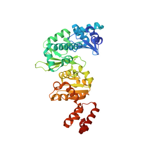
    The coordination of primase function within the replisome is an essential but poorly understood feature of lagging strand synthesis. By using crystallography and small-angle X-ray scattering (SAXS), we show that functional elements of bacterial primase transition between two dominant conformations: an extended form that uncouples a regulatory domain from its associated RNA polymerase core and a compact state that sequesters the regulatory region from the site of primer synthesis. FRET studies and priming assays reveal that the regulatory domain of one primase subunit productively associates with nucleic acid that is bound to the polymerase domain of a second protomer in trans. This intersubunit interaction allows primase to select initiation sites on template DNA and implicates the regulatory domain as a "molecular brake" that restricts primer length. Our data suggest that the replisome may cooperatively use multiple primases and this conformational switch to control initiation frequency, processivity, and ultimately, Okazaki fragment synthesis.


  • Organizational Affiliation

    Department of Molecular and Cell Biology, University of California, Berkeley, Berkeley, California 94720, USA.


Macromolecules
Find similar proteins by:  (by identity cutoff)  |  3D Structure
Entity ID: 1
MoleculeChains Sequence LengthOrganismDetailsImage
DNA primase407Aquifex aeolicusMutation(s): 0 
Gene Names: dnaG
EC: 2.7.7
UniProt
Find proteins for O67465 (Aquifex aeolicus (strain VF5))
Explore O67465 
Go to UniProtKB:  O67465
Entity Groups  
Sequence Clusters30% Identity50% Identity70% Identity90% Identity95% Identity100% Identity
UniProt GroupO67465
Sequence Annotations
Expand
  • Reference Sequence
Small Molecules
Ligands 1 Unique
IDChains Name / Formula / InChI Key2D Diagram3D Interactions
ZN
Query on ZN

Download Ideal Coordinates CCD File 
B [auth A]ZINC ION
Zn
PTFCDOFLOPIGGS-UHFFFAOYSA-N
Experimental Data & Validation

Experimental Data

  • Method: X-RAY DIFFRACTION
  • Resolution: 2.00 Å
  • R-Value Free: 0.238 
  • R-Value Work: 0.203 
  • R-Value Observed: 0.205 
  • Space Group: P 21 21 21
Unit Cell:
Length ( Å )Angle ( ˚ )
a = 43.222α = 90
b = 66.403β = 90
c = 138.157γ = 90
Software Package:
Software NamePurpose
REFMACrefinement
DENZOdata reduction
SCALEPACKdata scaling
AMoREphasing

Structure Validation

View Full Validation Report


Entry History 

Deposition Data

Revision History  (Full details and data files)

  • Version 1.0: 2005-11-15
    Type: Initial release
  • Version 1.1: 2008-05-01
    Changes: Version format compliance
  • Version 1.2: 2011-07-13
    Changes: Advisory, Version format compliance
  • Version 1.3: 2024-02-14
    Changes: Data collection, Database references, Derived calculations