Effects of disease-modifying anti-rheumatic drugs (DMARDs) on the activities of rheumatoid arthritis-associated cathepsins K and S.
Weidauer, E., Yasuda, Y., Biswal, B.K., Cherny, M., James, M.N., Bromme, D.(2007) Biol Chem 388: 331-336
- PubMed: 17338641 Search on PubMed
- DOI: https://doi.org/10.1515/BC.2007.037
- Primary Citation Related Structures:
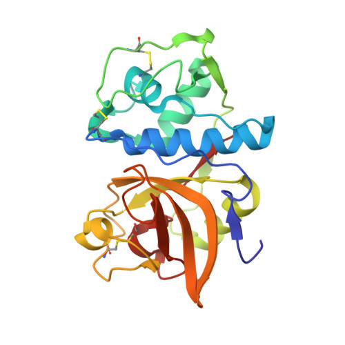
2ATO - PubMed Abstract:
Rheumatoid arthritis is an inflammatory and disabling joint disease affecting 0.5-1.5% of the population. Although various anti-inflammatory (NSAIDs) and disease-modifying (DMARDs) drugs are in clinical use, their precise mechanisms of action are not always defined. In this report, we discuss the effects of widely used DMARDs such as gold derivatives and chloroquine on cathepsins K and S, which have been implicated as critical mediators of inflammation and joint erosion in rheumatoid arthritis. We demonstrate that clinically potent gold derivatives inhibit cathepsins K and S in in vitro and cell-based assays. An X-ray analysis of the gold thiomalate/cathepsin K complex reveals that the inhibitor is bound to the active-site cysteine residue of the protease. Chloroquine, a lysosomotropic agent of lower clinical potency than gold derivatives, inhibits neutral pH-labile cathepsins intracellularly, but does not affect the neutral pH-stable cathepsin S. The potent inhibition of cathepsins implicated in the pathogenesis of rheumatoid arthritis by gold derivatives may explain the therapeutic efficacy of these drugs.
- Department of Human Genetics, Mount Sinai School of Medicine, New York, NY 10029, USA.
Organizational Affiliation: