2ARR

Human plasminogen activator inhibitor-2.[loop (66-98) deletion mutant] complexed with peptide n-acetyl-teaaagmggvmtgr-oh


Experimental Data Snapshot

  • Method: X-RAY DIFFRACTION
  • Resolution: 1.55 Å
  • R-Value Free: 0.220 
  • R-Value Work: 0.186 
  • R-Value Observed: 0.187 

wwPDB Validation   3D Report Full Report


This is version 1.5 of the entry. See complete history


Literature

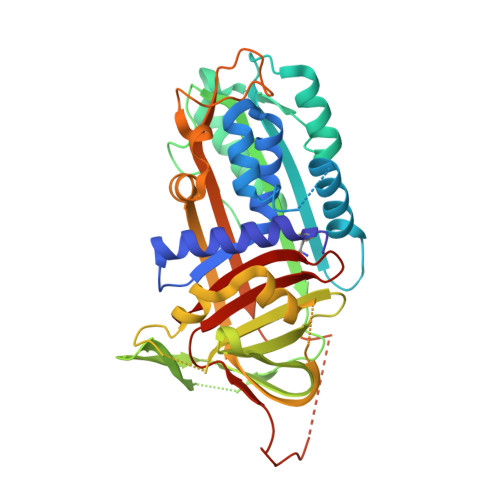
Plasminogen activator inhibitor-2 is highly tolerant to P8 residue substitution--implications for serpin mechanistic model and prediction of nsSNP activities

Di Giusto, D.A.Sutherland, A.P.Jankova, L.Harrop, S.J.Curmi, P.M.King, G.C.

(2005) J Mol Biol 353: 1069-1080

  • DOI: https://doi.org/10.1016/j.jmb.2005.09.008
  • Primary Citation of Related Structures:  
    2ARQ, 2ARR

  • PubMed Abstract: 

    The serine protease inhibitor (serpin) superfamily is involved in a wide range of cellular processes including fibrinolysis, angiogenesis, apoptosis, inflammation, metastasis and viral pathogenesis. Here, we investigate the unique mousetrap inhibition mechanism of serpins through saturation mutagenesis of the P8 residue for a typical family member, plasminogen activator inhibitor-2 (PAI-2). A number of studies have proposed an important role for the P8 residue in the efficient insertion and stabilisation of the cleaved reactive centre loop (RCL), which is a key event in the serpin inhibitory mechanism. The importance of this residue for inhibition of the PAI-2 protease target urinary plasminogen activator (urokinase, uPA) is confirmed, although a high degree of tolerance to P8 substitution is observed. Out of 19 possible PAI-2 P8 mutants, 16 display inhibitory activities within an order of magnitude of the wild-type P8 Thr species. Crystal structures of complexes between PAI-2 and RCL-mimicking peptides with P8 Met or Asp mutations are determined, and structural comparison with the wild-type complex substantiates the ability of the S8 pocket to accommodate disparate side-chains. These data indicate that the identity of the P8 residue is not a determinant of efficient RCL insertion, and provide further evidence for functional plasticity of key residues within enzyme structures. Poor correlation of observed PAI-2 P8 mutant activities with a range of physicochemical, evolutionary and thermodynamic predictive indices highlights the practical limitations of existing approaches to predicting the molecular phenotype of protein variants.


  • Organizational Affiliation

    School of Biotechnology and Biomolecular Sciences, The University of New South Wales, Sydney, NSW 2052, Australia.


Macromolecules
Find similar proteins by:  (by identity cutoff)  |  3D Structure
Entity ID: 1
MoleculeChains Sequence LengthOrganismDetailsImage
Plasminogen activator inhibitor-2382Homo sapiensMutation(s): 0 
UniProt & NIH Common Fund Data Resources
Find proteins for P05120 (Homo sapiens)
Explore P05120 
Go to UniProtKB:  P05120
PHAROS:  P05120
GTEx:  ENSG00000197632 
Entity Groups  
Sequence Clusters30% Identity50% Identity70% Identity90% Identity95% Identity100% Identity
UniProt GroupP05120
Sequence Annotations
Expand
  • Reference Sequence

Find similar proteins by:  Sequence   |   3D Structure  

Entity ID: 2
MoleculeChains Sequence LengthOrganismDetailsImage
14-mer from Plasminogen activator inhibitor-2B [auth P]15Homo sapiensMutation(s): 1 
UniProt & NIH Common Fund Data Resources
Find proteins for P05120 (Homo sapiens)
Explore P05120 
Go to UniProtKB:  P05120
PHAROS:  P05120
GTEx:  ENSG00000197632 
Entity Groups  
Sequence Clusters30% Identity50% Identity70% Identity90% Identity95% Identity100% Identity
UniProt GroupP05120
Sequence Annotations
Expand
  • Reference Sequence
Experimental Data & Validation

Experimental Data

  • Method: X-RAY DIFFRACTION
  • Resolution: 1.55 Å
  • R-Value Free: 0.220 
  • R-Value Work: 0.186 
  • R-Value Observed: 0.187 
  • Space Group: P 21 21 2
Unit Cell:
Length ( Å )Angle ( ˚ )
a = 91.952α = 90
b = 103.802β = 90
c = 41.406γ = 90
Software Package:
Software NamePurpose
REFMACrefinement
MOSFLMdata reduction
CCP4data scaling
AMoREphasing

Structure Validation

View Full Validation Report


Entry History 

Deposition Data

Revision History  (Full details and data files)

  • Version 1.0: 2006-07-11
    Type: Initial release
  • Version 1.1: 2008-04-30
    Changes: Version format compliance
  • Version 1.2: 2011-07-13
    Changes: Atomic model, Database references, Derived calculations, Non-polymer description, Structure summary, Version format compliance
  • Version 1.3: 2019-12-11
    Changes: Data collection, Database references, Derived calculations
  • Version 1.4: 2021-11-10
    Changes: Database references, Derived calculations
  • Version 1.5: 2023-10-25
    Changes: Data collection, Refinement description