2ARO

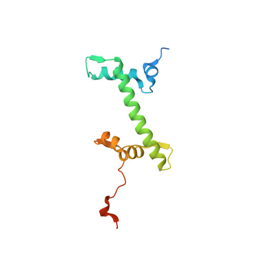
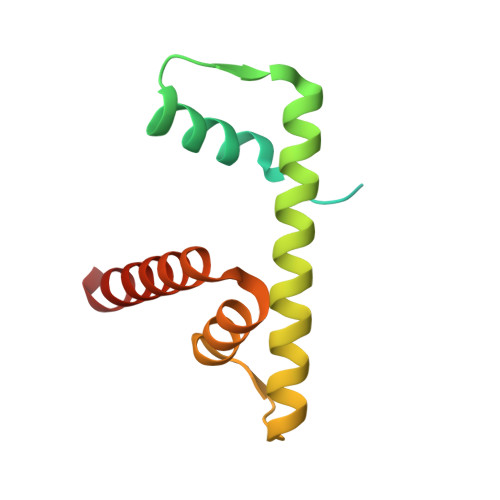
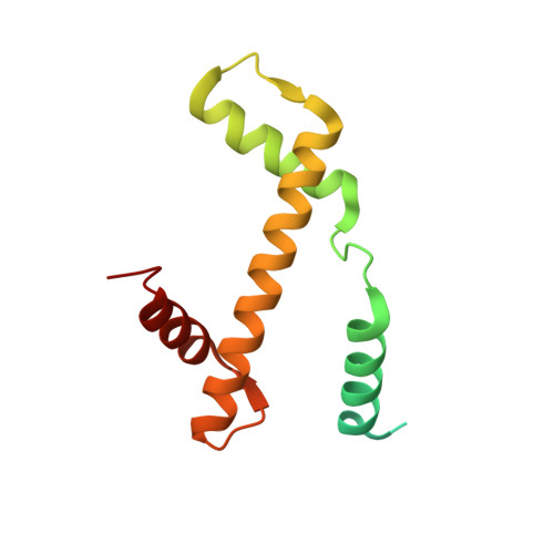
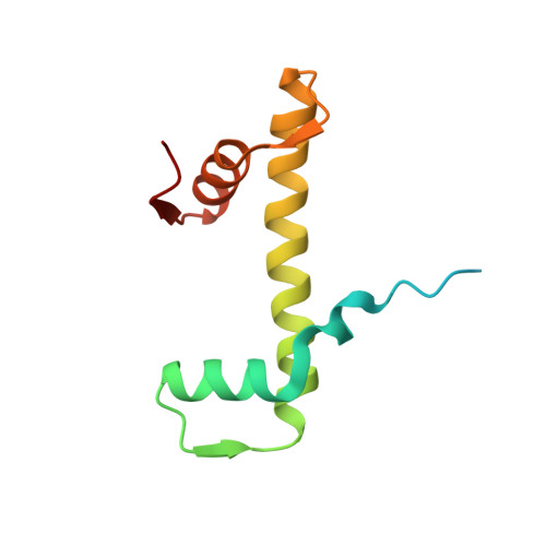
Crystal Structure Of The Native Histone Octamer To 2.1 Angstrom Resolution, Crystalised In The Presence Of S-Nitrosoglutathione


Experimental Data Snapshot

  • Method: X-RAY DIFFRACTION
  • Resolution: 2.10 Å
  • R-Value Free: 0.225 
  • R-Value Work: 0.184 
  • R-Value Observed: 0.186 

wwPDB Validation   3D Report Full Report


This is version 1.2 of the entry. See complete history


Literature

The oxidised histone octamer does not form a H3 disulphide bond.

Wood, C.M.Sodngam, S.Nicholson, J.M.Lambert, S.J.Reynolds, C.D.Baldwin, J.P.

(2006) Biochim Biophys Acta 1764: 1356-1362

  • DOI: 10.1016/j.bbapap.2006.06.014
  • Primary Citation of Related Structures:  
    2ARO

  • PubMed Abstract: 
  • A H3 dimer band is produced when purified native histone octamers are run on an SDS-PAGE gel in a beta-mercaptoethanol-free environment. To investigate this, native histone octamer crystals, derived from chicken erythrocytes, and of structure (H2A-H2B)-(H4-H3)-(H3'-H4')-(H2B'-H2A'), were grown in 2 M KCl, 1 ...

    A H3 dimer band is produced when purified native histone octamers are run on an SDS-PAGE gel in a beta-mercaptoethanol-free environment. To investigate this, native histone octamer crystals, derived from chicken erythrocytes, and of structure (H2A-H2B)-(H4-H3)-(H3'-H4')-(H2B'-H2A'), were grown in 2 M KCl, 1.35 M potassium phosphates and 250-350 microM of the oxidising agent S-nitrosoglutathione, pH 6.9. X-ray diffraction data were acquired to 2.10 A resolution, yielding a structure with an Rwork value of 18.6% and an Rfree of 22.5%. The space group is P6(5), the asymmetric unit of which contains one complete octamer. Compared to the 1.90 A resolution, unoxidised native histone octamer structure, the crystals show a reduction of 2.5% in the c-axis of the unit cell, and free-energy calculations reveal that the H3-H3' dimer interface in the latter has become thermodynamically stable, in contrast to the former. Although the inter-sulphur distance of the two H3 cysteines in the oxidised native histone octamer has reduced to 6 A from the 7 A of the unoxidised form, analysis of the hydrogen bonds that constitute the (H4-H3)-(H3'-H4') tetramer indicates that the formation of a disulphide bond in the H3-H3' dimer interface is incompatible with stable tetramer formation. The biochemical and biophysical evidence, taken as a whole, is indicative of crystals that have a stable H3-H3' dimer interface, possibly extending to the interface within an isolated H3-H3' dimer, observed in SDS-PAGE gels.


    Organizational Affiliation

    School of Biomolecular Sciences, Liverpool John Moores University, Byrom Street, Liverpool, L3 3AF, UK. c.m.wood@ljmu.ac.uk


Macromolecules
Find similar proteins by:  (by identity cutoff)  |  3D Structure
Entity ID: 1
MoleculeChainsSequence LengthOrganismDetailsImage
Histone H2A-IVA, E129Gallus gallusMutation(s): 0 
UniProt
Find proteins for P02263 (Gallus gallus)
Explore P02263 
Go to UniProtKB:  P02263
Entity Groups  
Sequence Clusters30% Identity50% Identity70% Identity90% Identity95% Identity100% Identity
UniProt GroupP02263
Protein Feature View
Expand
  • Reference Sequence
Find similar proteins by:  (by identity cutoff)  |  3D Structure
Entity ID: 2
MoleculeChainsSequence LengthOrganismDetailsImage
Histone H2BB, F126Gallus gallusMutation(s): 0 
Gene Names: H2B-IH2B-IIH2B-IIIH2B-IVH2B-VI
UniProt
Find proteins for P0C1H3 (Gallus gallus)
Explore P0C1H3 
Go to UniProtKB:  P0C1H3
Entity Groups  
Sequence Clusters30% Identity50% Identity70% Identity90% Identity95% Identity100% Identity
UniProt GroupP0C1H3
Protein Feature View
Expand
  • Reference Sequence
Find similar proteins by:  (by identity cutoff)  |  3D Structure
Entity ID: 3
MoleculeChainsSequence LengthOrganismDetailsImage
HISTONE H3C, G136Gallus gallusMutation(s): 0 
Gene Names: H3-IH3-IIH3-IIIH3-IVH3-VH3-VIH3-VIIH3-VIII
UniProt
Find proteins for P84229 (Gallus gallus)
Explore P84229 
Go to UniProtKB:  P84229
Entity Groups  
Sequence Clusters30% Identity50% Identity70% Identity90% Identity95% Identity100% Identity
UniProt GroupP84229
Protein Feature View
Expand
  • Reference Sequence
Find similar proteins by:  (by identity cutoff)  |  3D Structure
Entity ID: 4
MoleculeChainsSequence LengthOrganismDetailsImage
HISTONE H4-VID, H103Gallus gallusMutation(s): 0 
Gene Names: H4-IH4-IIH4-IIIH4-IVH4-VH4-VIH4-VII
UniProt
Find proteins for P62801 (Gallus gallus)
Explore P62801 
Go to UniProtKB:  P62801
Entity Groups  
Sequence Clusters30% Identity50% Identity70% Identity90% Identity95% Identity100% Identity
UniProt GroupP62801
Protein Feature View
Expand
  • Reference Sequence
Small Molecules
Ligands 2 Unique
IDChainsName / Formula / InChI Key2D Diagram3D Interactions
PO4
Query on PO4

Download Ideal Coordinates CCD File 
I [auth A],
O [auth B],
R [auth C],
Y [auth E],
Z [auth E]
PHOSPHATE ION
O4 P
NBIIXXVUZAFLBC-UHFFFAOYSA-K
 Ligand Interaction
CL
Query on CL

Download Ideal Coordinates CCD File 
AA [auth E],
BA [auth F],
CA [auth F],
DA [auth F],
EA [auth G],
AA [auth E],
BA [auth F],
CA [auth F],
DA [auth F],
EA [auth G],
FA [auth G],
GA [auth G],
HA [auth G],
IA [auth H],
J [auth A],
JA [auth H],
K [auth A],
L [auth A],
M [auth A],
N [auth A],
P [auth B],
Q [auth B],
S [auth C],
T [auth C],
U [auth C],
V [auth D],
W [auth D],
X [auth D]
CHLORIDE ION
Cl
VEXZGXHMUGYJMC-UHFFFAOYSA-M
 Ligand Interaction
Experimental Data & Validation

Experimental Data

  • Method: X-RAY DIFFRACTION
  • Resolution: 2.10 Å
  • R-Value Free: 0.225 
  • R-Value Work: 0.184 
  • R-Value Observed: 0.186 
  • Space Group: P 65
Unit Cell:
Length ( Å )Angle ( ˚ )
a = 158.084α = 90
b = 158.084β = 90
c = 101.037γ = 120
Software Package:
Software NamePurpose
REFMACrefinement
MOSFLMdata reduction

Structure Validation

View Full Validation Report


Entry History 

Deposition Data

Revision History  (Full details and data files)

  • Version 1.0: 2005-08-30
    Type: Initial release
  • Version 1.1: 2008-04-30
    Changes: Version format compliance
  • Version 1.2: 2011-07-13
    Changes: Advisory, Version format compliance