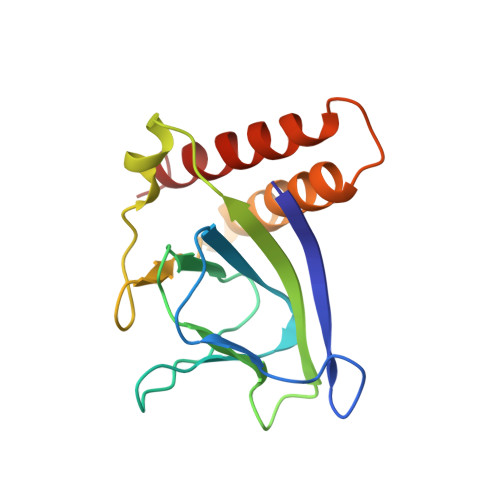
2ARA

APO FORM OF ESCHERICHIA COLI REGULATORY PROTEIN ARAC


Experimental Data Snapshot

  • Method: X-RAY DIFFRACTION
  • Resolution: 2.80 Å
  • R-Value Free: 0.337 
  • R-Value Work: 0.216 
  • R-Value Observed: 0.216 

wwPDB Validation   3D Report Full Report


This is version 1.3 of the entry. See complete history


Literature

Structural basis for ligand-regulated oligomerization of AraC.

Soisson, S.M.MacDougall-Shackleton, B.Schleif, R.Wolberger, C.

(1997) Science 276: 421-425

  • DOI: https://doi.org/10.1126/science.276.5311.421
  • Primary Citation of Related Structures:  
    2ARA, 2ARC

  • PubMed Abstract: 

    The crystal structure of the arabinose-binding and dimerization domain of the Escherchia coli gene regulatory protein AraC was determined in the presence and absence of L-arabinose. The 1.5 angstrom structure of the arabinose-bound molecule shows that the protein adopts an unusual fold, binding sugar within a beta barrel and completely burying the arabinose with the amino-terminal arm of the protein. Dimer contacts in the presence of arabinose are mediated by an antiparallel coiled-coil. In the 2.8 angstrom structure of the uncomplexed protein, the amino-terminal arm is disordered, uncovering the sugar-binding pocket and allowing it to serve as an oligomerization interface. The ligand-gated oligomerization as seen in AraC provides the basis of a plausible mechanism for modulating the protein's DNA-looping properties.


  • Organizational Affiliation

    Department of Biophysics and Biophysical Chemistry, Johns Hopkins University School of Medicine, Baltimore, MD 21205, USA.


Macromolecules
Find similar proteins by:  (by identity cutoff)  |  3D Structure
Entity ID: 1
MoleculeChains Sequence LengthOrganismDetailsImage
ARAC149Escherichia coliMutation(s): 0 
Gene Names: ARAC
UniProt
Find proteins for P0A9E0 (Escherichia coli (strain K12))
Explore P0A9E0 
Go to UniProtKB:  P0A9E0
Entity Groups  
Sequence Clusters30% Identity50% Identity70% Identity90% Identity95% Identity100% Identity
UniProt GroupP0A9E0
Sequence Annotations
Expand
  • Reference Sequence
Experimental Data & Validation

Experimental Data

  • Method: X-RAY DIFFRACTION
  • Resolution: 2.80 Å
  • R-Value Free: 0.337 
  • R-Value Work: 0.216 
  • R-Value Observed: 0.216 
  • Space Group: P 31 2 1
Unit Cell:
Length ( Å )Angle ( ˚ )
a = 57.37α = 90
b = 57.37β = 90
c = 83.45γ = 120
Software Package:
Software NamePurpose
DENZOdata reduction
SCALEPACKdata scaling
X-PLORmodel building
X-PLORrefinement
X-PLORphasing

Structure Validation

View Full Validation Report


Entry History 

Deposition Data

Revision History  (Full details and data files)

  • Version 1.0: 1997-05-15
    Type: Initial release
  • Version 1.1: 2008-03-24
    Changes: Version format compliance
  • Version 1.2: 2011-07-13
    Changes: Version format compliance
  • Version 1.3: 2023-08-09
    Changes: Database references, Refinement description