2AOX | pdb_00002aox

Histamine Methyltransferase (Primary Variant T105) Complexed with the Acetylcholinesterase Inhibitor and Altzheimer's Disease Drug Tacrine


Experimental Data Snapshot

  • Method: X-RAY DIFFRACTION
  • Resolution: 3.12 Å
  • R-Value Free: 
    0.285 (Depositor) 
  • R-Value Work: 
    0.229 (Depositor) 
  • R-Value Observed: 
    0.235 (Depositor) 

Starting Model: experimental
View more details

wwPDB Validation 3D Report Full Report

Validation slider image for 2AOX

This is version 1.3 of the entry. See complete history

Literature

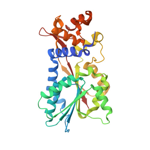
Structural basis for inhibition of histamine N-methyltransferase by diverse drugs

Horton, J.R.Sawada, K.Nishibori, M.Cheng, X.

(2005) J Mol Biology 353: 334-344

  • DOI: https://doi.org/10.1016/j.jmb.2005.08.040
  • Primary Citation Related Structures: 
    2AOT, 2AOU, 2AOV, 2AOW, 2AOX

  • PubMed Abstract: 

    In mammals, histamine action is terminated through metabolic inactivation by histamine N-methyltransferase (HNMT) and diamine oxidase. In addition to three well-studied pharmacological functions, smooth muscle contraction, increased vascular permeability, and stimulation of gastric acid secretion, histamine plays important roles in neurotransmission, immunomodulation, and regulation of cell proliferation. The histamine receptor H1 antagonist diphenhydramine, the antimalarial drug amodiaquine, the antifolate drug metoprine, and the anticholinesterase drug tacrine (an early drug for Alzheimer's disease) are surprisingly all potent HNMT inhibitors, having inhibition constants in the range of 10-100nM. We have determined the structural mode of interaction of these four inhibitors with HNMT. Despite their structural diversity, they all occupy the histamine-binding site, thus blocking access to the enzyme's active site. Near the N terminus of HNMT, several aromatic residues (Phe9, Tyr15, and Phe19) adopt different rotamer conformations or become disordered in the enzyme-inhibitor complexes, accommodating the diverse, rigid hydrophobic groups of the inhibitors. The maximized shape complementarity between the protein aromatic side-chains and aromatic ring(s) of the inhibitors are responsible for the tight binding of these varied inhibitors.


  • Organizational Affiliation
    • Department of Biochemistry Emory University School of Medicine, 1510 Clifton Road Atlanta, GA 30322, USA.

Macromolecule Content 

  • Total Structure Weight: 67.06 kDa 
  • Atom Count: 4,511 
  • Modeled Residue Count: 576 
  • Deposited Residue Count: 584 
  • Unique protein chains: 1

Macromolecules

Find similar proteins by:|  3D Structure
Entity ID: 1
MoleculeChains  Sequence LengthOrganismDetailsImage
Histamine N-methyltransferase
A, B
292Homo sapiensMutation(s): 0 
Gene Names: HNMT
EC: 2.1.1.8
UniProt & NIH Common Fund Data Resources
Find proteins for P50135 (Homo sapiens)
Explore P50135 
Go to UniProtKB:  P50135
PHAROS:  P50135
GTEx:  ENSG00000150540 
Entity Groups
Sequence Clusters30% Identity50% Identity70% Identity90% Identity95% Identity100% Identity
UniProt GroupP50135
Sequence Annotations
Expand
Reference Sequence

Small Molecules

Experimental Data & Validation

Experimental Data

  • Method: X-RAY DIFFRACTION
  • Resolution: 3.12 Å
  • R-Value Free:  0.285 (Depositor) 
  • R-Value Work:  0.229 (Depositor) 
  • R-Value Observed: 0.235 (Depositor) 
Space Group: P 6
Unit Cell:
Length ( Å )Angle ( ˚ )
a = 132.34α = 90
b = 132.34β = 90
c = 64.58γ = 120
Software Package:
Software NamePurpose
DENZOdata reduction
SCALEPACKdata scaling
GLRFphasing
CNSrefinement

Structure Validation

View Full Validation Report


Entry History 

Deposition Data

Revision History  (Full details and data files)

  • Version 1.0: 2005-09-13
    Type: Initial release
  • Version 1.1: 2008-04-30
    Changes: Version format compliance
  • Version 1.2: 2011-07-13
    Changes: Version format compliance
  • Version 1.3: 2023-08-23
    Changes: Data collection, Database references, Derived calculations, Refinement description