2AOQ | pdb_00002aoq

Crystal structure of MutH-unmethylated DNA complex


Experimental Data Snapshot

  • Method: X-RAY DIFFRACTION
  • Resolution: 2.20 Å
  • R-Value Free: 
    0.220 (Depositor), 0.223 (DCC) 
  • R-Value Work: 
    0.199 (Depositor), 0.199 (DCC) 
  • R-Value Observed: 
    0.199 (Depositor) 

Starting Model: experimental
View more details

wwPDB Validation 3D Report Full Report

Validation slider image for 2AOQ

This is version 1.3 of the entry. See complete history

Literature

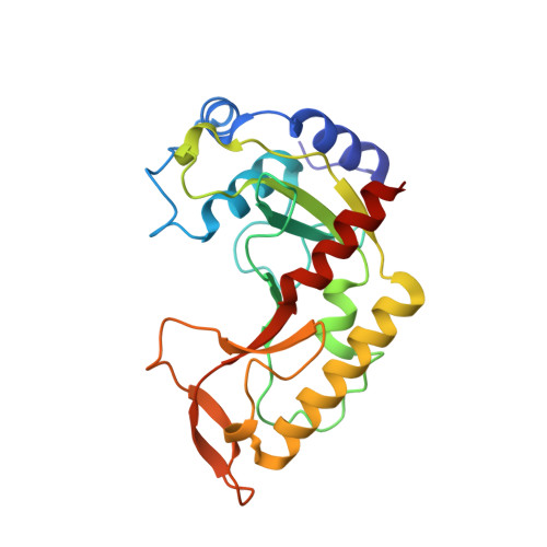
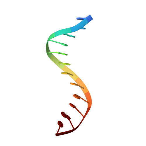
MutH complexed with hemi- and unmethylated DNAs: coupling base recognition and DNA cleavage.

Lee, J.Y.Chang, J.Joseph, N.Ghirlando, R.Rao, D.N.Yang, W.

(2005) Mol Cell 20: 155-166

  • DOI: https://doi.org/10.1016/j.molcel.2005.08.019
  • Primary Citation Related Structures: 
    2AOQ, 2AOR

  • PubMed Abstract: 

    MutH initiates mismatch repair by nicking the transiently unmethylated daughter strand 5' to a GATC sequence. Here, we report crystal structures of MutH complexed with hemimethylated and unmethylated GATC substrates. Both structures contain two Ca2+ ions jointly coordinated by a conserved aspartate and the scissile phosphate, as observed in the restriction endonucleases BamHI and BglI. In the hemimethylated complexes, the active site is more compact and DNA cleavage is more efficient. The Lys residue in the conserved DEK motif coordinates the nucleophilic water in conjunction with the phosphate 3' to the scissile bond; the same Lys is also hydrogen bonded with a carbonyl oxygen in the DNA binding module. We propose that this Lys, which is conserved in many restriction endonucleases and is replaced by Glu or Gln in BamHI and BglII, is a sensor for DNA binding and the linchpin that couples base recognition and DNA cleavage.


  • Organizational Affiliation
    • Laboratory of Molecular Biology, National Institute of Diabetes and Digestive and Kidney Diseases, National Institutes of Health, Bethesda, MD 20892, USA.

Macromolecule Content 

  • Total Structure Weight: 32.34 kDa 
  • Atom Count: 2,352 
  • Modeled Residue Count: 244 
  • Deposited Residue Count: 247 
  • Unique protein chains: 1
  • Unique nucleic acid chains: 1

Macromolecules


Find similar proteins by:|  3D Structure
Entity ID: 2
MoleculeChains  Sequence LengthOrganismDetailsImage
DNA mismatch repair protein mutHC [auth A]223Haemophilus influenzaeMutation(s): 0 
Gene Names: mutH
UniProt
Find proteins for P44688 (Haemophilus influenzae (strain ATCC 51907 / DSM 11121 / KW20 / Rd))
Explore P44688 
Go to UniProtKB:  P44688
Entity Groups
Sequence Clusters30% Identity50% Identity70% Identity90% Identity95% Identity100% Identity
UniProt GroupP44688
Sequence Annotations
Expand
Reference Sequence
Find similar nucleic acids by:  Sequence
Entity ID: 1
MoleculeChains LengthOrganismImage
5'-D(*GP*CP*AP*TP*GP*AP*TP*CP*AP*TP*GP*C)-3'A [auth B],
B [auth C]
12N/A
Sequence Annotations
Expand
Reference Sequence

Experimental Data & Validation

Experimental Data

  • Method: X-RAY DIFFRACTION
  • Resolution: 2.20 Å
  • R-Value Free:  0.220 (Depositor), 0.223 (DCC) 
  • R-Value Work:  0.199 (Depositor), 0.199 (DCC) 
  • R-Value Observed: 0.199 (Depositor) 
Space Group: P 63
Unit Cell:
Length ( Å )Angle ( ˚ )
a = 114.624α = 90
b = 114.624β = 90
c = 46.755γ = 120
Software Package:
Software NamePurpose
CNSrefinement
HKL-2000data reduction
SCALEPACKdata scaling
AMoREphasing

Structure Validation

View Full Validation Report


Entry History 

Deposition Data

Revision History  (Full details and data files)

  • Version 1.0: 2005-10-11
    Type: Initial release
  • Version 1.1: 2008-04-30
    Changes: Version format compliance
  • Version 1.2: 2011-07-13
    Changes: Version format compliance
  • Version 1.3: 2023-08-23
    Changes: Data collection, Database references, Derived calculations, Refinement description