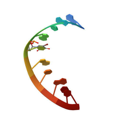
Structure of an RNA duplex r(GGCGBrUGCGCU)2 with terminal and internal tandem G.U base pairs.
Utsunomiya, R., Suto, K., Balasundaresan, D., Fukamizu, A., Kumar, P.K., Mizuno, H.(2006) Acta Crystallogr D Biol Crystallogr 62: 331-338
- PubMed: 16510980 Search on PubMed
- DOI: https://doi.org/10.1107/S0907444905043210
- Primary Citation Related Structures:
2AO5 - PubMed Abstract:
The crystal structure of a self-complementary RNA duplex r(GGCG(Br)UGCGCU)(2) with terminal G.U and internal tandem G.U base pairs has been determined at 2.1 Angstroms resolution. The crystals belong to the tetragonal space group P4(3), with unit-cell parameters a = b = 37.69, c = 96.28 Angstroms and two duplexes in the asymmetric unit. The two strands of each duplex are related by a pseudodyad axis. The structure was refined to final R(work) and R(free) values of 20.9 and 25.3%, respectively. The duplexes stack in an end-to-end manner, forming infinite columns along the c axis. This is the first structural study of an RNA duplex containing G.U pairs at the termini. The stacking overlaps of the terminal G.U base pairs with their adjacent Watson-Crick base pairs are larger than those of Watson-Crick base pairs of the 5'-YR-3'/3'-RY-5' type. The terminal G.U base pairs of neighbouring duplexes are also stacked with each other. An alternating underwound-overwound pattern of the twist angles is seen at each step along the duplex. This observation is typical for internal tandem G.U pairs, while the terminal G.U base pairs exhibit high twist angles with the adjacent Watson-Crick pairs. The 3'-side of U of the internal G.U base pair, which is unstacked, appears to be stabilized by pi-cation interaction with an Mg(2+) ion.
- Institute of Applied Biochemistry, University of Tsukuba, Ibaraki, Japan.
Organizational Affiliation: