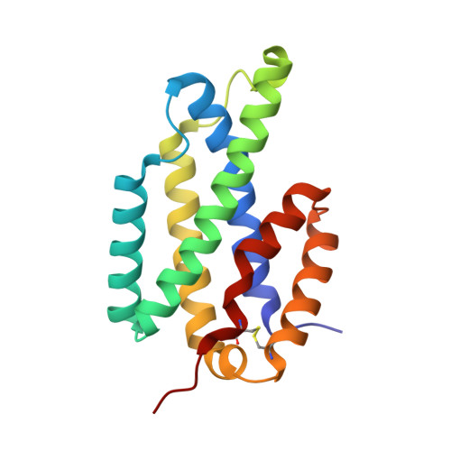
The 2.15 A crystal structure of Mycobacterium tuberculosis chorismate mutase reveals an unexpected gene duplication and suggests a role in host-pathogen interactions.
Qamra, R., Prakash, P., Aruna, B., Hasnain, S.E., Mande, S.C.(2006) Biochemistry 45: 6997-7005
- PubMed: 16752890
- DOI: https://doi.org/10.1021/bi0606445
- Primary Citation of Related Structures:
2AO2 - PubMed Abstract:
Chorismate mutase catalyzes the first committed step toward the biosynthesis of the aromatic amino acids, phenylalanine and tyrosine. While this biosynthetic pathway exists exclusively in the cell cytoplasm, the Mycobacterium tuberculosis enzyme has been shown to be secreted into the extracellular medium. The secretory nature of the enzyme and its existence in M. tuberculosis as a duplicated gene are suggestive of its role in host-pathogen interactions. We report here the crystal structure of homodimeric chorismate mutase (Rv1885c) from M. tuberculosis determined at 2.15 A resolution. The structure suggests possible gene duplication within each subunit of the dimer (residues 35-119 and 130-199) and reveals an interesting proline-rich region on the protein surface (residues 119-130), which might act as a recognition site for protein-protein interactions. The structure also offers an explanation for its regulation by small ligands, such as tryptophan, a feature previously unknown in the prototypical Escherichia coli chorismate mutase. The tryptophan ligand is found to be sandwiched between the two monomers in a dimer contacting residues 66-68. The active site in the "gene-duplicated" monomer is occupied by a sulfate ion and is located in the first half of the polypeptide, unlike in the Saccharomyces cerevisiae (yeast) enzyme, where it is located in the later half. We hypothesize that the M. tuberculosis chorismate mutase might have a role to play in host-pathogen interactions, making it an important target for designing inhibitor molecules against the deadly pathogen.
- Department of Biophysics, University of Delhi South Campus, New Delhi, India.
Organizational Affiliation:


