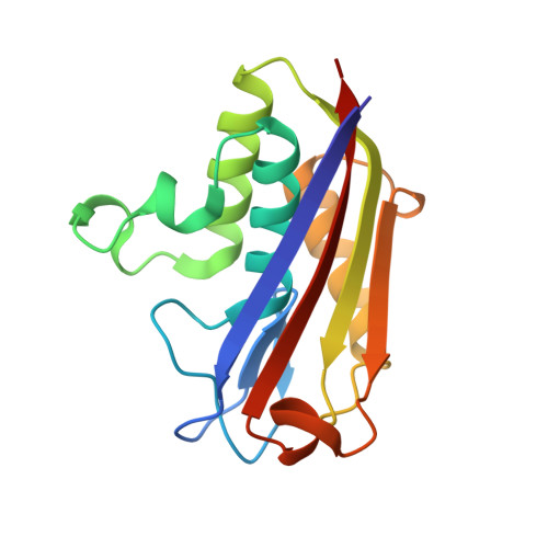
2AMT

Structure of 2C-Methyl-D-Erythritol 2,4-Clycodiphosphate Synthase complexed with a CDP derived fluorescent inhibitor


Experimental Data Snapshot

  • Method: X-RAY DIFFRACTION
  • Resolution: 2.30 Å
  • R-Value Free: 0.280 
  • R-Value Work: 0.243 
  • R-Value Observed: 0.245 

wwPDB Validation   3D Report Full Report


Ligand Structure Quality Assessment 


This is version 1.2 of the entry. See complete history


Literature

Fluorescent Inhibitors for IspF, an Enzyme in the Non-Mevalonate Pathway for Isoprenoid Biosynthesis and a Potential Target for Antimalarial Therapy.

Crane, C.M.Kaiser, J.Ramsden, N.L.Lauw, S.Rohdich, F.Eisenreich, W.Hunter, W.N.Bacher, A.Diederich, F.

(2006) Angew Chem Int Ed Engl 45: 1069-1074


Macromolecules
Find similar proteins by:  (by identity cutoff)  |  3D Structure
Entity ID: 1
MoleculeChainsSequence LengthOrganismDetailsImage
2-C-methyl-D-erythritol 2,4-cyclodiphosphate synthase
A, B, C, D, E, F
A, B, C, D, E, F
159Escherichia coliMutation(s): 0 
Gene Names: ispFmecS
EC: 4.6.1.12
UniProt
Find proteins for P62617 (Escherichia coli (strain K12))
Explore P62617 
Go to UniProtKB:  P62617
Entity Groups  
Sequence Clusters30% Identity50% Identity70% Identity90% Identity95% Identity100% Identity
UniProt GroupP62617
Protein Feature View
Expand
  • Reference Sequence
Small Molecules
Ligands 3 Unique
IDChainsName / Formula / InChI Key2D Diagram3D Interactions
1AA
Query on 1AA

Download Ideal Coordinates CCD File 
H [auth A],
L [auth C],
M [auth C],
O [auth D],
R [auth E],
H [auth A],
L [auth C],
M [auth C],
O [auth D],
R [auth E],
S [auth E]
5'-O-[({[(2-{[(2-AMINOPHENYL)CARBONYL]OXY}ETHYL)OXY]PHOSPHINATO}OXY)PHOSPHINATO]CYTIDINE
C18 H24 N4 O13 P2
PGXVRIOOHRVKLC-DTZQCDIJSA-N
 Ligand Interaction
GPP
Query on GPP

Download Ideal Coordinates CCD File 
I [auth A],
P [auth D]
GERANYL DIPHOSPHATE
C10 H20 O7 P2
GVVPGTZRZFNKDS-JXMROGBWSA-N
 Ligand Interaction
ZN
Query on ZN

Download Ideal Coordinates CCD File 
G [auth A],
J [auth B],
K [auth C],
N [auth D],
Q [auth E],
G [auth A],
J [auth B],
K [auth C],
N [auth D],
Q [auth E],
T [auth F]
ZINC ION
Zn
PTFCDOFLOPIGGS-UHFFFAOYSA-N
 Ligand Interaction
Binding Affinity Annotations 
IDSourceBinding Affinity
1AA PDBBind:  2AMT Kd: 3.60e+4 (nM) from 1 assay(s)
Binding MOAD:  2AMT Kd: 3.60e+4 (nM) from 1 assay(s)
Experimental Data & Validation

Experimental Data

  • Method: X-RAY DIFFRACTION
  • Resolution: 2.30 Å
  • R-Value Free: 0.280 
  • R-Value Work: 0.243 
  • R-Value Observed: 0.245 
  • Space Group: P 1 21 1
Unit Cell:
Length ( Å )Angle ( ˚ )
a = 54.033α = 90
b = 115.269β = 90.18
c = 87.609γ = 90
Software Package:
Software NamePurpose
REFMACrefinement
MOSFLMdata reduction
CCP4data scaling
MOLREPphasing

Structure Validation

View Full Validation Report


Ligand Structure Quality Assessment 


Entry History 

Deposition Data

Revision History  (Full details and data files)

  • Version 1.0: 2006-02-28
    Type: Initial release
  • Version 1.1: 2008-04-30
    Changes: Version format compliance
  • Version 1.2: 2011-07-13
    Changes: Version format compliance