2AK3

THE THREE-DIMENSIONAL STRUCTURE OF THE COMPLEX BETWEEN MITOCHONDRIAL MATRIX ADENYLATE KINASE AND ITS SUBSTRATE AMP AT 1.85 ANGSTROMS RESOLUTION


Experimental Data Snapshot

  • Method: X-RAY DIFFRACTION
  • Resolution: 1.85 Å
  • R-Value Work: 0.189 
  • R-Value Observed: 0.189 

wwPDB Validation   3D Report Full Report


This is version 1.3 of the entry. See complete history


Literature

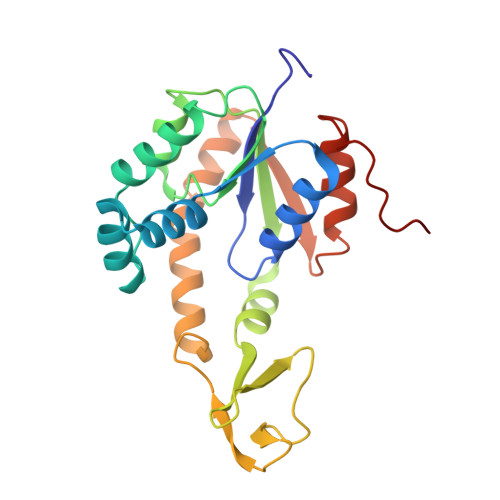
The refined structure of the complex between adenylate kinase from beef heart mitochondrial matrix and its substrate AMP at 1.85 A resolution.

Diederichs, K.Schulz, G.E.

(1991) J Mol Biol 217: 541-549

  • DOI: https://doi.org/10.1016/0022-2836(91)90756-v
  • Primary Citation of Related Structures:  
    2AK3

  • PubMed Abstract: 

    The crystal structure of the complex between adenylate kinase from bovine mitochondrial matrix and its substrate AMP has been refined at 1.85 A resolution (1 A = 0.1 nm). Based on 42,519 independent reflections of better than 10 A resolution, a final R-factor of 18.9% was obtained with a model obeying standard geometry within 0.016 A in bond lengths and 3.2 degrees in bond angles. There are two enzyme: substrate complexes in the asymmetric unit, each consisting of 226 amino acid residues, one AMP and one sulfate ion. A superposition of the two full-length polypeptides revealed deviations that can be described as small relative movements of three domains. Best superpositions of individual domains yielded a residual overall root-mean-square deviation of 0.3 A for the backbone atoms and 0.5 A for the sidechains. The final model contains 381 solvent molecules in the asymmetric unit, 2 x 72 = 144 of which occupy corresponding positions in both complexes.


  • Organizational Affiliation

    Institut für Organische Chemie und Biochemie Freiburg, F.R.G.


Macromolecules
Find similar proteins by:  (by identity cutoff)  |  3D Structure
Entity ID: 1
MoleculeChains Sequence LengthOrganismDetailsImage
ADENYLATE KINASE ISOENZYME-3
A, B
226Bos taurusMutation(s): 0 
EC: 2.7.4.10
UniProt
Find proteins for P08760 (Bos taurus)
Explore P08760 
Go to UniProtKB:  P08760
Entity Groups  
Sequence Clusters30% Identity50% Identity70% Identity90% Identity95% Identity100% Identity
UniProt GroupP08760
Sequence Annotations
Expand
  • Reference Sequence
Small Molecules
Experimental Data & Validation

Experimental Data

  • Method: X-RAY DIFFRACTION
  • Resolution: 1.85 Å
  • R-Value Work: 0.189 
  • R-Value Observed: 0.189 
  • Space Group: P 21 21 21
Unit Cell:
Length ( Å )Angle ( ˚ )
a = 49.83α = 90
b = 66.99β = 90
c = 155.41γ = 90
Software Package:
Software NamePurpose
X-PLORmodel building
X-PLORrefinement
X-PLORphasing

Structure Validation

View Full Validation Report


Entry History 

Deposition Data

Revision History  (Full details and data files)

  • Version 1.0: 1995-05-08
    Type: Initial release
  • Version 1.1: 2008-03-24
    Changes: Version format compliance
  • Version 1.2: 2011-07-13
    Changes: Version format compliance
  • Version 1.3: 2024-02-14
    Changes: Data collection, Database references, Derived calculations, Other