2AIZ | pdb_00002aiz

Solution structure of peptidoglycan associated lipoprotein from Haemophilus influenza bound to UDP-N-acetylmuramoyl-L-alanyl-D-glutamyl-meso-2,6-diaminopimeloyl-D-alanyl-D-alanine


Experimental Data Snapshot

  • Method: SOLUTION NMR
  • Conformers Calculated: 100 
  • Conformers Submitted: 20 
  • Selection Criteria: structures with the least restraint violations 

wwPDB Validation 3D Report Full Report

Validation slider image for 2AIZ

This is version 2.1 of the entry. See complete history

Literature

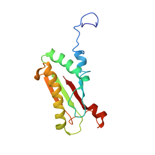
Peptidoglycan recognition by pal, an outer membrane lipoprotein.

Parsons, L.M.Lin, F.Orban, J.

(2006) Biochemistry 45: 2122-2128

  • DOI: https://doi.org/10.1021/bi052227i
  • Primary Citation Related Structures: 
    2AIZ

  • PubMed Abstract: 

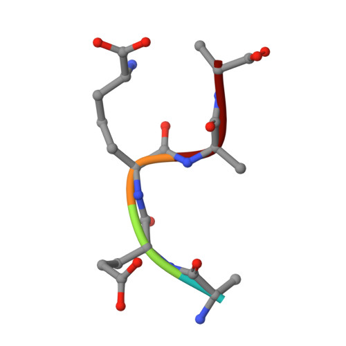
    Peptidoglycan-associated lipoprotein (Pal) is a potential vaccine candidate from Haemophilus influenzae that is highly conserved in Gram-negative bacteria and anchored to the outer membrane through an N-terminal lipid attachment. Pal stabilizes the outer membrane by providing a noncovalent link to the peptidoglycan (PG) layer through a periplasmic domain. Using NMR spectroscopy, we determined the three-dimensional structure of a complex between the periplasmic domain of Pal and a biosynthetic peptidoglycan precursor (PG-P), UDP-N-acetylmuramyl-L-Ala-alpha-d-Glu-m-Dap-D-Ala-d-Ala (m-Dap is meso-diaminopimelate). Pal has a binding pocket lined with conserved surface residues that interacts exclusively with the peptide portion of the ligand. The m-Dap residue, which is mainly found in the cell walls of Gram-negative bacteria, is sequestered in this pocket and plays an important role by forming hydrogen bond and hydrophobic contacts to Pal. The structure provides insight into the mode of cell wall recognition for a broad class of Gram-negative membrane proteins, including OmpA and MotB, which have peptidoglycan-binding domains homologous to that of Pal.


  • Organizational Affiliation
    • Center for Advanced Research in Biotechnology, University of Maryland Biotechnology Institute, 9600 Gudelsky Drive, Rockville, Maryland 20850, USA.

Macromolecule Content 

  • Total Structure Weight: 15.48 kDa 
  • Atom Count: 1,086 
  • Modeled Residue Count: 139 
  • Deposited Residue Count: 139 
  • Unique protein chains: 2

Macromolecules

Find similar proteins by:|  3D Structure
Entity ID: 1
MoleculeChains  Sequence LengthOrganismDetailsImage
Outer membrane protein P6 (Fragment)A [auth P]134Haemophilus influenzaeMutation(s): 0 
Gene Names: ompP6
UniProt
Find proteins for P10324 (Haemophilus influenzae (strain ATCC 51907 / DSM 11121 / KW20 / Rd))
Explore P10324 
Go to UniProtKB:  P10324
Entity Groups
Sequence Clusters30% Identity50% Identity70% Identity90% Identity95% Identity100% Identity
UniProt GroupP10324
Sequence Annotations
Expand
Reference Sequence
Find similar proteins by:  Sequence   |   3D Structure  
Entity ID: 2
MoleculeChains  Sequence LengthOrganismDetailsImage
L-alanyl-D-glutamyl-meso-2,6-diaminopimeloyl-D-alanyl-D-alanineB [auth U]5synthetic constructMutation(s): 0 

Small Molecules

Ligands 2 Unique
IDChains Name / Formula / InChI Key2D Diagram3D Interactions
UDP

Query on UDP


Download:Ideal Coordinates CCD File
D [auth U]URIDINE-5'-DIPHOSPHATE
C9 H14 N2 O12 P2
XCCTYIAWTASOJW-XVFCMESISA-N
AMU

Query on AMU


Download:Ideal Coordinates CCD File
C [auth U]N-acetyl-beta-muramic acid
C11 H19 N O8
MNLRQHMNZILYPY-YVNCZSHWSA-N
Modified Residues  1 Unique
IDChains TypeFormula2D DiagramParent
6CL
Query on 6CL
B [auth U]L-PEPTIDE LINKINGC7 H15 N2 O4LYS
DAL
Query on DAL
B [auth U]D-PEPTIDE LINKINGC3 H7 N O2

--

DGL
Query on DGL
B [auth U]D-PEPTIDE LINKINGC5 H9 N O4

--

Experimental Data & Validation

Experimental Data

  • Method: SOLUTION NMR
  • Conformers Calculated: 100 
  • Conformers Submitted: 20 
  • Selection Criteria: structures with the least restraint violations 

Structure Validation

View Full Validation Report


Entry History 

Deposition Data

Revision History  (Full details and data files)

  • Version 1.0: 2006-03-14
    Type: Initial release
  • Version 1.1: 2008-04-30
    Changes: Version format compliance
  • Version 1.2: 2011-07-13
    Changes: Version format compliance
  • Version 1.3: 2020-07-29
    Type: Remediation
    Reason: Carbohydrate remediation
    Changes: Data collection, Derived calculations, Structure summary
  • Version 2.0: 2023-02-15
    Changes: Atomic model, Data collection, Database references, Derived calculations, Polymer sequence, Source and taxonomy, Structure summary
  • Version 2.1: 2024-10-30
    Changes: Data collection, Structure summary