2AEW

A model for growth hormone receptor activation based on subunit rotation within a receptor dimer


Experimental Data Snapshot

  • Method: X-RAY DIFFRACTION
  • Resolution: 2.70 Å
  • R-Value Free: 0.302 
  • R-Value Work: 0.257 
  • R-Value Observed: 0.262 

wwPDB Validation   3D Report Full Report


This is version 1.4 of the entry. See complete history


Literature

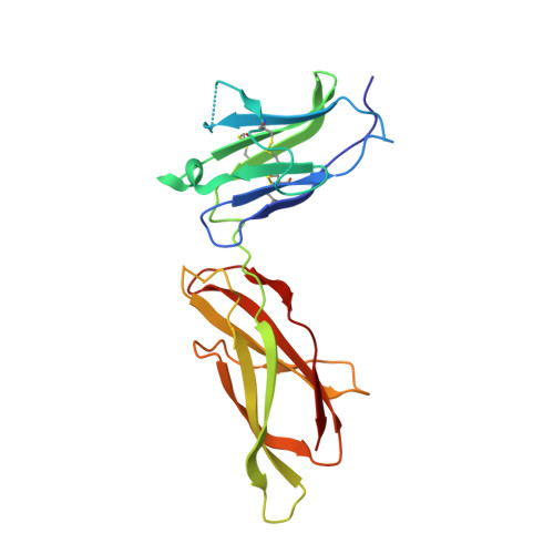
Model for growth hormone receptor activation based on subunit rotation within a receptor dimer.

Brown, R.J.Adams, J.J.Pelekanos, R.A.Wan, Y.McKinstry, W.J.Palethorpe, K.Seeber, R.M.Monks, T.A.Eidne, K.A.Parker, M.W.Waters, M.J.

(2005) Nat Struct Mol Biol 12: 814-821

  • DOI: https://doi.org/10.1038/nsmb977
  • Primary Citation of Related Structures:  
    2AEW

  • PubMed Abstract: 

    Growth hormone is believed to activate the growth hormone receptor (GHR) by dimerizing two identical receptor subunits, leading to activation of JAK2 kinase associated with the cytoplasmic domain. However, we have reported previously that dimerization alone is insufficient to activate full-length GHR. By comparing the crystal structure of the liganded and unliganded human GHR extracellular domain, we show here that there is no substantial change in its conformation on ligand binding. However, the receptor can be activated by rotation without ligand by inserting a defined number of alanine residues within the transmembrane domain. Fluorescence resonance energy transfer (FRET), bioluminescence resonance energy transfer (BRET) and coimmunoprecipitation studies suggest that receptor subunits undergo specific transmembrane interactions independent of hormone binding. We propose an activation mechanism involving a relative rotation of subunits within a dimeric receptor as a result of asymmetric placement of the receptor-binding sites on the ligand.


  • Organizational Affiliation

    Institute for Molecular Bioscience and School of Biomedical Sciences, University of Queensland, St. Lucia, Queensland, Australia.


Macromolecules
Find similar proteins by:  (by identity cutoff)  |  3D Structure
Entity ID: 1
MoleculeChains Sequence LengthOrganismDetailsImage
Growth hormone receptor
A, B
205Homo sapiensMutation(s): 0 
UniProt & NIH Common Fund Data Resources
Find proteins for P10912 (Homo sapiens)
Explore P10912 
Go to UniProtKB:  P10912
PHAROS:  P10912
GTEx:  ENSG00000112964 
Entity Groups  
Sequence Clusters30% Identity50% Identity70% Identity90% Identity95% Identity100% Identity
UniProt GroupP10912
Sequence Annotations
Expand
  • Reference Sequence
Experimental Data & Validation

Experimental Data

  • Method: X-RAY DIFFRACTION
  • Resolution: 2.70 Å
  • R-Value Free: 0.302 
  • R-Value Work: 0.257 
  • R-Value Observed: 0.262 
  • Space Group: C 2 2 21
Unit Cell:
Length ( Å )Angle ( ˚ )
a = 99.72α = 90
b = 112.167β = 90
c = 93.16γ = 90
Software Package:
Software NamePurpose
DENZOdata reduction
SCALEPACKdata scaling
AMoREphasing
CNSrefinement
PDB_EXTRACTdata extraction

Structure Validation

View Full Validation Report


Entry History 

Deposition Data

Revision History  (Full details and data files)

  • Version 1.0: 2005-11-01
    Type: Initial release
  • Version 1.1: 2007-10-16
    Changes: Version format compliance
  • Version 1.2: 2011-07-13
    Changes: Version format compliance
  • Version 1.3: 2017-10-11
    Changes: Refinement description
  • Version 1.4: 2023-10-25
    Changes: Data collection, Database references, Refinement description