2ADO

Crystal Structure Of The Brct Repeat Region From The Mediator of DNA damage checkpoint protein 1, MDC1


Experimental Data Snapshot

  • Method: X-RAY DIFFRACTION
  • Resolution: 1.45 Å
  • R-Value Free: 0.212 
  • R-Value Work: 0.195 

wwPDB Validation   3D Report Full Report


This is version 1.4 of the entry. See complete history


Literature

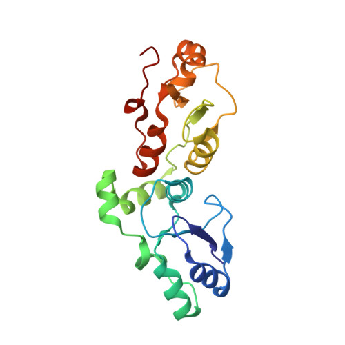
Structure of the BRCT Repeat Domain of MDC1 and Its Specificity for the Free COOH-terminal End of the {gamma}-H2AX Histone Tail.

Lee, M.S.Edwards, R.A.Thede, G.L.Glover, J.N.

(2005) J Biol Chem 280: 32053-32056

  • DOI: https://doi.org/10.1074/jbc.C500273200
  • Primary Citation of Related Structures:  
    2ADO

  • PubMed Abstract: 

    MDC1 (mediator of DNA damage checkpoint protein 1) regulates the recognition and repair of DNA double strand breaks in mammalian cells through its interactions with nuclear foci containing the COOH-terminally phosphorylated form of the histone variant, H2AX. Here we demonstrate that the tandem BRCT repeats of MDC1 directly bind to the phosphorylated tail of H2AX-Ser(P)-Gln-Glu-Tyr, in a manner that is critically dependent on the free carboxylate group of the COOH-terminal Tyr residue. We have determined the x-ray crystal structure of the MDC1 BRCT repeats at 1.45 Angstroms resolution. By a comparison with the structure of the BRCA1 BRCT bound to a phosphopeptide, we suggest that two arginine residues in MDC1, Arg(1932) and Arg(1933) may recognize the COOH terminus of the peptide as well as the penultimate Glu of H2AX, while Gln(2013) may provide additional specificity for the COOH-terminal Tyr.


  • Organizational Affiliation

    Department of Biochemistry, University of Alberta, Edmonton, Canada.


Macromolecules
Find similar proteins by:  (by identity cutoff)  |  3D Structure
Entity ID: 1
MoleculeChains Sequence LengthOrganismDetailsImage
Mediator of DNA damage checkpoint protein 1
A, B
196Homo sapiensMutation(s): 0 
Gene Names: MDC1KIAA0170NFBD1
UniProt & NIH Common Fund Data Resources
Find proteins for Q14676 (Homo sapiens)
Explore Q14676 
Go to UniProtKB:  Q14676
PHAROS:  Q14676
GTEx:  ENSG00000137337 
Entity Groups  
Sequence Clusters30% Identity50% Identity70% Identity90% Identity95% Identity100% Identity
UniProt GroupQ14676
Sequence Annotations
Expand
  • Reference Sequence
Experimental Data & Validation

Experimental Data

  • Method: X-RAY DIFFRACTION
  • Resolution: 1.45 Å
  • R-Value Free: 0.212 
  • R-Value Work: 0.195 
  • Space Group: P 1
Unit Cell:
Length ( Å )Angle ( ˚ )
a = 42.051α = 72.95
b = 44.44β = 87.54
c = 61.942γ = 61.8
Software Package:
Software NamePurpose
DENZOdata reduction
SCALEPACKdata scaling
SOLVEphasing
RESOLVEphasing
CNSrefinement
PDB_EXTRACTdata extraction

Structure Validation

View Full Validation Report


Entry History 

Deposition Data

Revision History  (Full details and data files)

  • Version 1.0: 2005-08-02
    Type: Initial release
  • Version 1.1: 2008-04-30
    Changes: Version format compliance
  • Version 1.2: 2011-07-13
    Changes: Version format compliance
  • Version 1.3: 2017-10-11
    Changes: Refinement description
  • Version 1.4: 2024-02-14
    Changes: Data collection, Database references