2ACJ

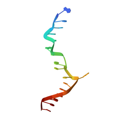
Crystal structure of the B/Z junction containing DNA bound to Z-DNA binding proteins


Experimental Data Snapshot

  • Method: X-RAY DIFFRACTION
  • Resolution: 2.60 Å
  • R-Value Free: 0.285 
  • R-Value Work: 0.238 
  • R-Value Observed: 0.243 

wwPDB Validation   3D Report Full Report


This is version 1.3 of the entry. See complete history


Literature

Crystal structure of a junction between B-DNA and Z-DNA reveals two extruded bases.

Ha, S.C.Lowenhaupt, K.Rich, A.Kim, Y.G.Kim, K.K.

(2005) Nature 437: 1183-1186

  • DOI: https://doi.org/10.1038/nature04088
  • Primary Citation of Related Structures:  
    2ACJ

  • PubMed Abstract: 

    Left-handed Z-DNA is a higher-energy form of the double helix, stabilized by negative supercoiling generated by transcription or unwrapping nucleosomes. Regions near the transcription start site frequently contain sequence motifs favourable for forming Z-DNA, and formation of Z-DNA near the promoter region stimulates transcription. Z-DNA is also stabilized by specific protein binding; several proteins have been identified with low nanomolar binding constants. Z-DNA occurs in a dynamic state, forming as a result of physiological processes then relaxing to the right-handed B-DNA. Each time a DNA segment turns into Z-DNA, two B-Z junctions form. These have been examined extensively, but their structure was unknown. Here we describe the structure of a B-Z junction as revealed by X-ray crystallography at 2.6 A resolution. A 15-base-pair segment of DNA is stabilized at one end in the Z conformation by Z-DNA binding proteins, while the other end remains B-DNA. Continuous stacking of bases between B-DNA and Z-DNA segments is found, with the breaking of one base pair at the junction and extrusion of the bases on each side (Fig. 1). These extruded bases may be sites for DNA modification.


  • Organizational Affiliation

    Department of Molecular Cell Biology, Samsung Biomedical Research Institute, Sungkyunkwan University School of Medicine, Suwon 440-746, Korea.


Macromolecules

Find similar proteins by:  (by identity cutoff)  |  3D Structure
Entity ID: 3
MoleculeChains Sequence LengthOrganismDetailsImage
Double-stranded RNA-specific adenosine deaminaseC [auth A],
D [auth B],
E [auth C],
F [auth D]
66Homo sapiensMutation(s): 0 
Gene Names: ADAR1
EC: 3.5.4 (PDB Primary Data), 3.5.4.37 (UniProt)
UniProt & NIH Common Fund Data Resources
Find proteins for P55265 (Homo sapiens)
Explore P55265 
Go to UniProtKB:  P55265
PHAROS:  P55265
GTEx:  ENSG00000160710 
Entity Groups  
Sequence Clusters30% Identity50% Identity70% Identity90% Identity95% Identity100% Identity
UniProt GroupP55265
Sequence Annotations
Expand
  • Reference Sequence

Find similar nucleic acids by:  Sequence   |   3D Structure  

Entity ID: 1
MoleculeChains LengthOrganismImage
5'-D(*GP*TP*CP*GP*CP*GP*CP*GP*CP*CP*AP*TP*AP*AP*AP*CP*C)-3'A [auth E]17N/A
Sequence Annotations
Expand
  • Reference Sequence

Find similar nucleic acids by:  Sequence   |   3D Structure  

Entity ID: 2
MoleculeChains LengthOrganismImage
5'-D(*AP*CP*GP*GP*TP*TP*TP*AP*TP*GP*GP*CP*GP*CP*GP*CP*G)-3'B [auth F]17N/A
Sequence Annotations
Expand
  • Reference Sequence
Experimental Data & Validation

Experimental Data

  • Method: X-RAY DIFFRACTION
  • Resolution: 2.60 Å
  • R-Value Free: 0.285 
  • R-Value Work: 0.238 
  • R-Value Observed: 0.243 
  • Space Group: P 61
Unit Cell:
Length ( Å )Angle ( ˚ )
a = 110.765α = 90
b = 110.765β = 90
c = 61.762γ = 120
Software Package:
Software NamePurpose
REFMACrefinement
HKL-2000data reduction
SCALEPACKdata scaling
SOLVEphasing

Structure Validation

View Full Validation Report


Entry History 

Deposition Data

Revision History  (Full details and data files)

  • Version 1.0: 2005-10-25
    Type: Initial release
  • Version 1.1: 2008-04-30
    Changes: Version format compliance
  • Version 1.2: 2011-07-13
    Changes: Advisory, Version format compliance
  • Version 1.3: 2024-03-13
    Changes: Data collection, Database references