2ACH

CRYSTAL STRUCTURE OF CLEAVED HUMAN ALPHA1-ANTICHYMOTRYPSIN AT 2.7 ANGSTROMS RESOLUTION AND ITS COMPARISON WITH OTHER SERPINS


Experimental Data Snapshot

  • Method: X-RAY DIFFRACTION
  • Resolution: 2.70 Å
  • R-Value Work: 0.181 

wwPDB Validation   3D Report Full Report


This is version 2.0 of the entry. See complete history


Literature

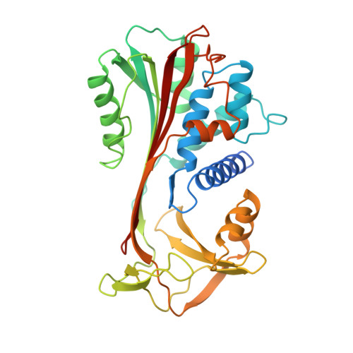
Crystal structure of cleaved human alpha 1-antichymotrypsin at 2.7 A resolution and its comparison with other serpins.

Baumann, U.Huber, R.Bode, W.Grosse, D.Lesjak, M.Laurell, C.B.

(1991) J Mol Biol 218: 595-606

  • DOI: https://doi.org/10.1016/0022-2836(91)90704-a
  • Primary Citation of Related Structures:  
    2ACH

  • PubMed Abstract: 

    The crystal structure of proteolytically modified human alpha 1-antichymotrypsin (ACT), a member of the serpin superfamily, has been solved by Paterson search techniques and refined to an R-factor of 18.0% at 2.7 A resolution with mean deviations from standard bond lengths and angles of 0.013 A and 3.1 degrees, respectively. The final model consists of 374 amino acid residues, 126 solvent molecules and five sugar residues. Asn70 could be identified unambiguously as a glycosylation site and Asn104 is probably also glycosylated. The structure of cleaved ACT is compared with cleaved alpha 1-antitrypsin (alpha 1 PI) and with plakalbumin, which are prototypical models for cleaved and intact serpins, respectively. Cleaved ACT is very similar to cleaved alpha 1 PI; in particular, it has strand s4A, which is liberated by proteolysis, inserted as the middle strand in beta-sheet A. ACT and alpha 1 PI differ locally only at sites of insertions, except at the segment s3C-turn-s4C, which is displaced by several angström units. This region of ACT is involved in DNA binding.


  • Organizational Affiliation

    Max-Planck-Institut für Biochemie, Martinsried, Germany.


Macromolecules
Find similar proteins by:  (by identity cutoff)  |  3D Structure
Entity ID: 1
MoleculeChains Sequence LengthOrganismDetailsImage
ALPHA 1-ANTICHYMOTRYPSIN360Homo sapiensMutation(s): 0 
UniProt & NIH Common Fund Data Resources
Find proteins for P01011 (Homo sapiens)
Explore P01011 
Go to UniProtKB:  P01011
PHAROS:  P01011
GTEx:  ENSG00000196136 
Entity Groups  
Sequence Clusters30% Identity50% Identity70% Identity90% Identity95% Identity100% Identity
UniProt GroupP01011
Sequence Annotations
Expand
  • Reference Sequence
Find similar proteins by:  (by identity cutoff)  |  3D Structure
Entity ID: 2
MoleculeChains Sequence LengthOrganismDetailsImage
ALPHA 1-ANTICHYMOTRYPSIN40Homo sapiensMutation(s): 0 
UniProt & NIH Common Fund Data Resources
Find proteins for P01011 (Homo sapiens)
Explore P01011 
Go to UniProtKB:  P01011
PHAROS:  P01011
GTEx:  ENSG00000196136 
Entity Groups  
Sequence Clusters30% Identity50% Identity70% Identity90% Identity95% Identity100% Identity
UniProt GroupP01011
Sequence Annotations
Expand
  • Reference Sequence
Oligosaccharides

Help

Entity ID: 3
MoleculeChains Length2D Diagram Glycosylation3D Interactions
beta-D-mannopyranose-(1-4)-2-acetamido-2-deoxy-beta-D-glucopyranose-(1-4)-2-acetamido-2-deoxy-beta-D-glucopyranose
C
3N-Glycosylation
Glycosylation Resources
GlyTouCan:  G15407YE
GlyCosmos:  G15407YE
GlyGen:  G15407YE
Entity ID: 4
MoleculeChains Length2D Diagram Glycosylation3D Interactions
2-acetamido-2-deoxy-beta-D-glucopyranose-(1-4)-2-acetamido-2-deoxy-beta-D-glucopyranose
D
2N-Glycosylation
Glycosylation Resources
GlyTouCan:  G42666HT
GlyCosmos:  G42666HT
GlyGen:  G42666HT
Small Molecules
Ligands 1 Unique
IDChains Name / Formula / InChI Key2D Diagram3D Interactions
PO4
Query on PO4

Download Ideal Coordinates CCD File 
E [auth A]PHOSPHATE ION
O4 P
NBIIXXVUZAFLBC-UHFFFAOYSA-K
Experimental Data & Validation

Experimental Data

  • Method: X-RAY DIFFRACTION
  • Resolution: 2.70 Å
  • R-Value Work: 0.181 
  • Space Group: P 41 21 2
Unit Cell:
Length ( Å )Angle ( ˚ )
a = 75.3α = 90
b = 75.3β = 90
c = 208γ = 90
Software Package:
Software NamePurpose
X-PLORrefinement

Structure Validation

View Full Validation Report


Entry History 

Deposition Data

Revision History  (Full details and data files)

  • Version 1.0: 1993-07-15
    Type: Initial release
  • Version 1.1: 2008-03-24
    Changes: Version format compliance
  • Version 1.2: 2011-07-13
    Changes: Non-polymer description, Version format compliance
  • Version 2.0: 2020-07-29
    Type: Remediation
    Reason: Carbohydrate remediation
    Changes: Advisory, Atomic model, Data collection, Derived calculations, Other, Structure summary