2ABK | pdb_00002abk

REFINEMENT OF THE NATIVE STRUCTURE OF ENDONUCLEASE III TO A RESOLUTION OF 1.85 ANGSTROM


Experimental Data Snapshot

  • Method: X-RAY DIFFRACTION
  • Resolution: 1.85 Å
  • R-Value Free: 
    0.216 (Depositor) 
  • R-Value Work: 
    0.185 (Depositor) 
  • R-Value Observed: 
    0.185 (Depositor) 

wwPDB Validation 3D Report Full Report

Validation slider image for 2ABK

This is version 1.4 of the entry. See complete history

Literature

Novel DNA binding motifs in the DNA repair enzyme endonuclease III crystal structure.

Thayer, M.M.Ahern, H.Xing, D.Cunningham, R.P.Tainer, J.A.

(1995) EMBO J 14: 4108-4120

  • DOI: https://doi.org/10.1002/j.1460-2075.1995.tb00083.x
  • Primary Citation Related Structures: 
    2ABK

  • PubMed Abstract: 

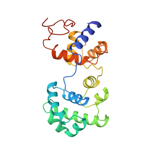
    The 1.85 A crystal structure of endonuclease III, combined with mutational analysis, suggests the structural basis for the DNA binding and catalytic activity of the enzyme. Helix-hairpin-helix (HhH) and [4Fe-4S] cluster loop (FCL) motifs, which we have named for their secondary structure, bracket the cleft separating the two alpha-helical domains of the enzyme. These two novel DNA binding motifs and the solvent-filled pocket in the cleft between them all lie within a positively charged and sequence-conserved surface region. Lys120 and Asp138, both shown by mutagenesis to be catalytically important, lie at the mouth of this pocket, suggesting that this pocket is part of the active site. The positions of the HhH motif and protruding FCL motif, which contains the DNA binding residue Lys191, can accommodate B-form DNA, with a flipped-out base bound within the active site pocket. The identification of HhH and FCL sequence patterns in other DNA binding proteins suggests that these motifs may be a recurrent structural theme for DNA binding proteins.


  • Organizational Affiliation
    • Scripps Research Institute, Department of Molecular Biology-MB4, La Jolla, CA 92037, USA.

Macromolecule Content 

  • Total Structure Weight: 23.95 kDa 
  • Atom Count: 1,798 
  • Modeled Residue Count: 211 
  • Deposited Residue Count: 211 
  • Unique protein chains: 1

Macromolecules

Find similar proteins by:|  3D Structure
Entity ID: 1
MoleculeChains  Sequence LengthOrganismDetailsImage
ENDONUCLEASE III211Escherichia coliMutation(s): 0 
EC: 4.2.99.18
UniProt
Find proteins for P0AB83 (Escherichia coli (strain K12))
Explore P0AB83 
Go to UniProtKB:  P0AB83
Entity Groups
Sequence Clusters30% Identity50% Identity70% Identity90% Identity95% Identity100% Identity
UniProt GroupP0AB83
Sequence Annotations
Expand
Reference Sequence

Small Molecules

Ligands 1 Unique
IDChains Name / Formula / InChI Key2D Diagram3D Interactions
SF4

Query on SF4


Download:Ideal Coordinates CCD File
B [auth A]IRON/SULFUR CLUSTER
Fe4 S4
LJBDFODJNLIPKO-UHFFFAOYSA-N

Experimental Data & Validation

Experimental Data

  • Method: X-RAY DIFFRACTION
  • Resolution: 1.85 Å
  • R-Value Free:  0.216 (Depositor) 
  • R-Value Work:  0.185 (Depositor) 
  • R-Value Observed: 0.185 (Depositor) 
Space Group: P 21 21 21
Unit Cell:
Length ( Å )Angle ( ˚ )
a = 48.5α = 90
b = 65.8β = 90
c = 86.8γ = 90
Software Package:
Software NamePurpose
X-PLORmodel building
X-PLORrefinement
XENGENdata reduction
X-PLORphasing

Structure Validation

View Full Validation Report


Entry History 

Deposition Data

Revision History  (Full details and data files)

  • Version 1.0: 1995-10-15
    Type: Initial release
  • Version 1.1: 2008-03-03
    Changes: Version format compliance
  • Version 1.2: 2011-07-13
    Changes: Version format compliance
  • Version 1.3: 2017-11-29
    Changes: Derived calculations, Other
  • Version 1.4: 2024-02-14
    Changes: Data collection, Database references, Derived calculations