Plasmodium falciparum glutathione S-transferase--structural and mechanistic studies on ligand binding and enzyme inhibition.
Hiller, N., Fritz-Wolf, K., Deponte, M., Wende, W., Zimmermann, H., Becker, K.(2006) Protein Sci 15: 281-289
- PubMed: 16385005 Search on PubMedSearch on PubMed Central
- DOI: https://doi.org/10.1110/ps.051891106
- Primary Citation Related Structures:
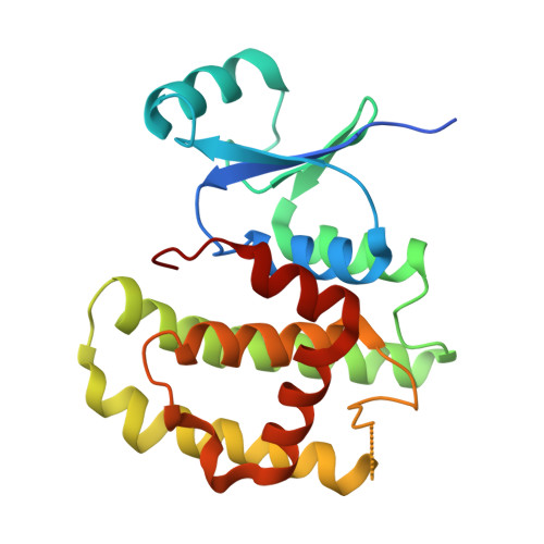
2AAW - PubMed Abstract:
Glutathione S-transferase of the malarial parasite Plasmodium falciparum (PfGST) represents a novel class of GST isoenzymes. Since the architecture of the PfGST substrate binding site differs significantly from its human counterparts and there is only this one isoenzyme present in the parasite, PfGST is considered a highly attractive target for antimalarial drug development. Here we report the mechanistic, kinetic, and structural characterization of PfGST as well as its interaction with different ligands. Our data indicate that in solution PfGST is present as a tetramer that dissociates into dimers in the presence of glutathione (GSH). Fluorescence spectroscopy shows that in the presence of GSH GST serves as ligandin for parasitotoxic ferriprotoporphyrin IX with a high- and a low-affinity binding site. This is supported by a clear uncompetitive inhibition type. Site-directed mutagenesis studies demonstrate that neither Cys 86 nor Cys 101 contribute to the peroxidase activity of the enzyme, which is thus performed GSH-dependently at the active site. Tyr 9 is responsible for the deprotonation of GSH and Lys 15, but also Gln 71 are involved in GSH binding. We furthermore report the 2.4 A resolution X-ray structure of PfGST cocrystallized with the inhibitor S-hexylglutathione. In comparison with a previously reported structure obtained by crystal soaking, differences occur at the C-terminal end of helix alpha4 and at the S-hexylmoiety of the inhibitor. We furthermore show that, in contrast to previous reports, the antimalarial drug artemisinin is not metabolized by PfGST.
- Interdisciplinary Research Center, Justus-Liebig-University, Heinrich-Buff-Ring 26-32, 35392 Giessen, Germany.
Organizational Affiliation: