Structures of the G81A mutant form of the active chimera of (S)-mandelate dehydrogenase and its complex with two of its substrates
Sukumar, N., Dewanti, A., Merli, A., Rossi, G.L., Mitra, B., Mathews, F.S.(2009) Acta Crystallogr D Biol Crystallogr 65: 543-552
- PubMed: 19465768 Search on PubMedSearch on PubMed Central
- DOI: https://doi.org/10.1107/S0907444909010270
- Primary Citation Related Structures:
2A7N, 2A7P, 2A85, 3GIY - PubMed Abstract:
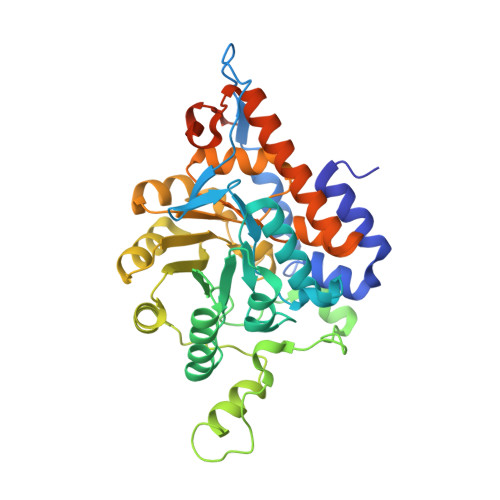
(S)-Mandelate dehydrogenase (MDH) from Pseudomonas putida, a membrane-associated flavoenzyme, catalyzes the oxidation of (S)-mandelate to benzoylformate. Previously, the structure of a catalytically similar chimera, MDH-GOX2, rendered soluble by the replacement of its membrane-binding segment with the corresponding segment of glycolate oxidase (GOX), was determined and found to be highly similar to that of GOX except within the substituted segments. Subsequent attempts to cocrystallize MDH-GOX2 with substrate proved unsuccessful. However, the G81A mutants of MDH and of MDH-GOX2 displayed approximately 100-fold lower reactivity with substrate and a modestly higher reactivity towards molecular oxygen. In order to understand the effect of the mutation and to identify the mode of substrate binding in MDH-GOX2, a crystallographic investigation of the G81A mutant of the MDH-GOX2 enzyme was initiated. The structures of ligand-free G81A mutant MDH-GOX2 and of its complexes with the substrates 2-hydroxyoctanoate and 2-hydroxy-3-indolelactate were determined at 1.6, 2.5 and 2.2 A resolution, respectively. In the ligand-free G81A mutant protein, a sulfate anion previously found at the active site is displaced by the alanine side chain introduced by the mutation. 2-Hydroxyoctanoate binds in an apparently productive mode for subsequent reaction, while 2-hydroxy-3-indolelactate is bound to the enzyme in an apparently unproductive mode. The results of this investigation suggest that a lowering of the polarity of the flavin environment resulting from the displacement of nearby water molecules caused by the glycine-to-alanine mutation may account for the lowered catalytic activity of the mutant enzyme, which is consistent with the 30 mV lower flavin redox potential. Furthermore, the altered binding mode of the indolelactate substrate may account for its reduced activity compared with octanoate, as observed in the crystalline state.
- NE-CAT and Department of Chemistry and Chemical Biology, Cornell University, Argonne National Laboratory, Argonne, IL 60439, USA.
Organizational Affiliation: