The Mitomycin C (MMC)-binding Protein from MMC-producing Microorganisms Protects from the Lethal Effect of Bleomycin: Crystallographic Analysis to Elucidate the Binding Mode of the Antibiotic to the Protein
Danshiitsoodol, N., de Pinho, C.A., Matoba, Y., Kumagai, T., Sugiyama, M.(2006) J Mol Biol 360: 398-408
- PubMed: 16756991 Search on PubMed
- DOI: https://doi.org/10.1016/j.jmb.2006.05.017
- Primary Citation Related Structures:
2A4W, 2A4X - PubMed Abstract:
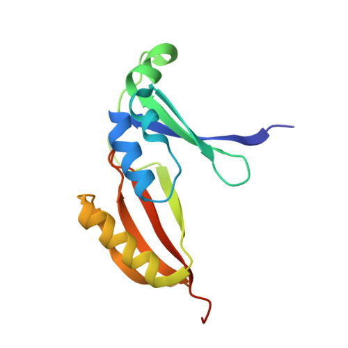
Antibiotic-producing microorganisms must be protected from the lethal effect of their own antibiotic. We have previously determined the X-ray crystal structure of the bleomycin (Bm)-binding protein, designated BLMA, as a self-resistance determinant from Bm-producing Streptomyces verticillus, which suggests that the binding of the first Bm to one of two pockets formed in the BLMA homodimer induces the cooperative binding of the second Bm to the other pocket. In the present study, we noticed that the X-ray crystallographic structure of a self-resistance determinant from a mitomycin C-producing microorganism, designated MRDP, reveals similarity to the folding pattern on the BLMA, although no sequence homology exists. To clarify the hypothesis that MRDP may function as a resistance determinant to Bm, we characterized and determined the crystal structure of MRDP complexed with the Cu(II)-bound form of BmA(2) grouped into the Bm family of antibiotics. The biochemical and structural studies for Bm binding provide evidence that the first Bm binds anti-cooperatively to a pocket of MRDP with binding affinity of the nanomolar order, whereas the second Bm binds to the other pocket, which has binding affinity of the micromolar order. The invisibility of the second Bm in the structure agrees with the observation that Escherichia coli-expressing MRDP displays lower resistance to Bm than that expressing BLMA. The structure of MRDP, which is complexed with the Cu(II)-bound BmA(2), revealed that the gamma-aminopropyldimethylsulphonium moiety of the antibiotic is sandwiched between the peripheral residues of the binding pocket and that its positively charged sulphonium head is accommodated completely in the negatively charged region of the MRDP pocket. Furthermore, the Cu(II)-bound BmA(2) has a very compact structure, in which the bithiazole ring of BmA(2) is folded back to the metal-binding domain.
- Department of Molecular Microbiology and Biotechnology, Graduate School of Biomedical Sciences, Hiroshima University, Kasumi 1-2-3, Minami-ku, Hiroshima 734-8551, Japan.
Organizational Affiliation: