Structural basis for sequential cleavage of fibrinopeptides upon fibrin assembly.
Pechik, I., Yakovlev, S., Mosesson, M.W., Gilliland, G.L., Medved, L.(2006) Biochemistry 45: 3588-3597
- PubMed: 16533041 Search on PubMedSearch on PubMed Central
- DOI: https://doi.org/10.1021/bi0525369
- Primary Citation Related Structures:
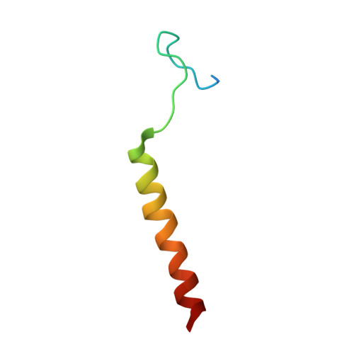
2A45 - PubMed Abstract:
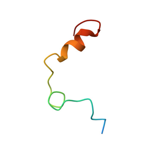
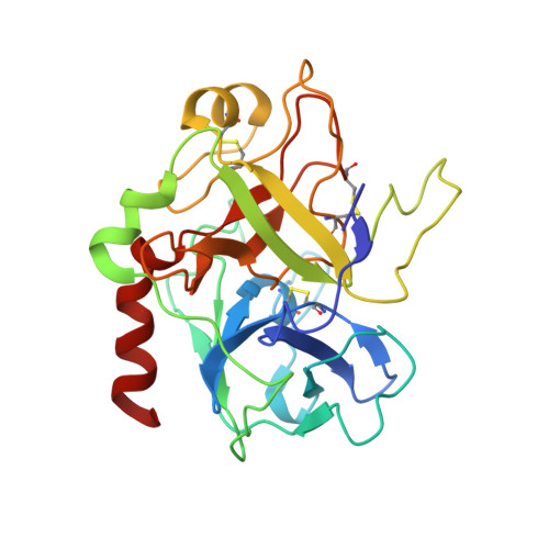
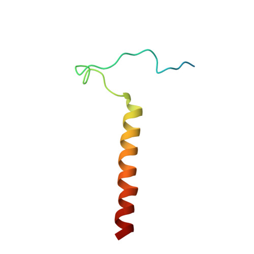
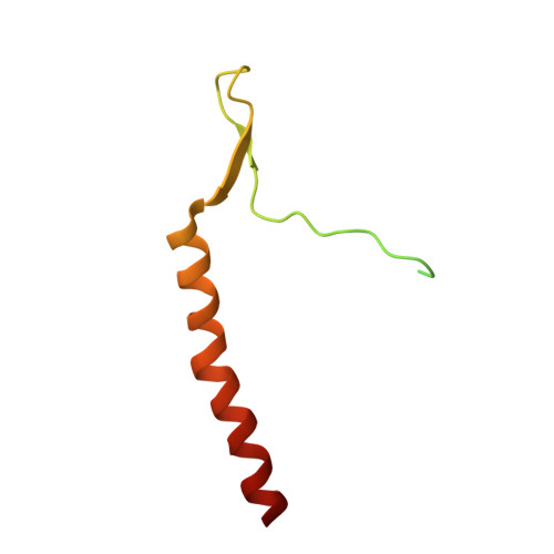
Nonsubstrate interaction of thrombin with fibrinogen promotes sequential cleavage of fibrinopeptides A and B (fpA and fpB, respectively) from the latter, resulting in its conversion into fibrin. The recently established crystal structure of human thrombin in complex with the central part of human fibrin clarified the mechanism of this interaction. Here, we reveal new details of the structure and present the results of molecular modeling of the fpA- and fpB-containing portions of the Aalpha and Bbeta chains, not identified in the complex, in both fibrinogen and protofibrils. The analysis of the results reveals that in fibrinogen the fpA-containing portions are in a more favorable position to bind in the active site cleft of bound thrombin. Surface plasmon resonance experiments establish that the fpB-containing portions interact with the fibrin-derived dimeric D-D fragment, suggesting that in protofibrils they bind to the newly formed DD regions bringing fpB into the vicinity of bound thrombin. These findings provide a coherent rationale for the preferential removal of fpA from fibrinogen at the first stage of fibrin assembly and the accelerated cleavage of fpB from protofibrils and/or fibrils at the second stage.
- Center for Advanced Research in Biotechnology, University of Maryland Biotechnology Institute and National Institute of Standards and Technology, Rockville, Maryland 20850, USA.
Organizational Affiliation: