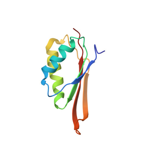
Solution structure, stability, and nucleic Acid binding of the hyperthermophile protein sso10b2.
Biyani, K., Kahsai, M.A., Clark, A.T., Armstrong, T.L., Edmondson, S.P., Shriver, J.W.(2005) Biochemistry 44: 14217-14230
- PubMed: 16245938 Search on PubMed
- DOI: https://doi.org/10.1021/bi051266r
- Primary Citation Related Structures:
2A2Y - PubMed Abstract:
The Sso10b (or Alba) family of proteins is a conserved group of archaeal and eukaryotic proteins which are thought to play a role in both chromatin organization and RNA metabolism. We describe here the solution structure and properties of Sso10b2 from Sulfolobus solfataricus. NMR data including residual dipolar couplings and (15)N relaxation data demonstrated that the protein adopts a beta(1)alpha(1)beta(2)alpha(2)beta(3)beta(4) topology with an IF-3-like fold. The protein dimerizes in solution at 30 degrees C via a hydrophobic surface defined by the C-terminal alpha(2)beta(3)beta(4) elements with a structure similar to one of the putative dimers indicated by previous crystal structures. DSC and circular dichroism data demonstrated an unusual two-state structural transition near the growth temperature which led to an increase in beta-sheet content without dissociation of the dimer. The cooperativity of the transition exceeded that of a dimer at pH 7, demonstrating the presence of higher order oligomers near the growth temperature at pH 7. Reverse titrations of Sso10b2 with nucleic acid showed that the protein binds single-stranded DNA (K(d) of 3 x 10(-)(7) M) with higher affinity than RNA (1.3 x 10(-)(6) M) or double-stranded DNA (1.5 x 10(-)(5) M) in 10 mM KH(2)PO(4) (pH 7.0, 20 degrees C). NMR chemical shift perturbation data indicated that single-stranded DNA and RNA binding occurred across the same dimer interface and encompassed a surface defined by the C-terminal ends of the beta(1), beta(2), and beta(3) strands of each monomer.
- Laboratory for Structural Biology, Department of Chemistry, Graduate Program in Biotechnology and Bioengineering, University of Alabama in Huntsville, Huntsville, Alabama 35899, USA.
Organizational Affiliation: