2A2A | pdb_00002a2a

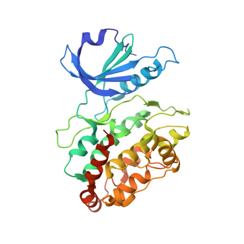
High-resolution crystallographic analysis of the autoinhibited conformation of a human death-associated protein kinase


Experimental Data Snapshot

  • Method: X-RAY DIFFRACTION
  • Resolution: 1.47 Å
  • R-Value Free: 
    0.207 (Depositor), 0.212 (DCC) 
  • R-Value Work: 
    0.150 (Depositor), 0.153 (DCC) 
  • R-Value Observed: 
    0.150 (Depositor) 

Starting Model: experimental
View more details

wwPDB Validation 3D Report Full Report

Validation slider image for 2A2A

This is version 1.4 of the entry. See complete history. 

Literature

Death-Associated Protein Kinase Activity Is Regulated by Coupled Calcium/Calmodulin Binding to Two Distinct Sites

Simon, B., Huart, A.S., Temmerman, K., Vahokoski, J., Mertens, H.D., Komadina, D., Hoffmann, J.E., Yumerefendi, H., Svergun, D.I., Kursula, P., Schultz, C., McCarthy, A.A., Hart, D.J., Wilmanns, M.

(2016) Structure 

  • DOI: https://doi.org/10.1016/j.str.2016.03.020
  • Primary Citation Related Structures: 
    1YRP, 2A2A, 2XZS

  • PubMed Abstract: 

    The regulation of many protein kinases by binding to calcium/calmodulin connects two principal mechanisms in signaling processes: protein phosphorylation and responses to dose- and time-dependent calcium signals. We used the calcium/calmodulin-dependent members of the death-associated protein kinase (DAPK) family to investigate the role of a basic DAPK signature loop near the kinase active site. In DAPK2, this loop comprises a novel dimerization-regulated calcium/calmodulin-binding site, in addition to a well-established calcium/calmodulin site in the C-terminal autoregulatory domain. Unexpectedly, impairment of the basic loop interaction site completely abolishes calcium/calmodulin binding and DAPK2 activity is reduced to a residual level, indicative of coupled binding to the two sites. This contrasts with the generally accepted view that kinase calcium/calmodulin interactions are autonomous of the kinase catalytic domain. Our data establish an intricate model of multi-step kinase activation and expand our understanding of how calcium binding connects with other mechanisms involved in kinase activity regulation.


  • Organizational Affiliation: 
    • European Molecular Biology Laboratory, Hamburg Unit, Notkestrasse 85, 22607 Hamburg, Germany.

Macromolecule Content 

  • Total Structure Weight: 149.85 kDa 
  • Atom Count: 11,556 
  • Modeled Residue Count: 1,213 
  • Deposited Residue Count: 1,284 
  • Unique protein chains: 1

Macromolecules

Find similar proteins by:|  3D Structure
Entity ID: 1
MoleculeChains  Sequence LengthOrganismDetailsImage
Death-associated protein kinase 2
A, B, C, D
321Homo sapiensMutation(s): 0 
EC: 2.7.1.37 (PDB Primary Data), 2.7.11.1 (UniProt)
UniProt & NIH Common Fund Data Resources
Find proteins for Q9UIK4 (Homo sapiens)
Explore Q9UIK4 
Go to UniProtKB:  Q9UIK4
PHAROS:  Q9UIK4
GTEx:  ENSG00000035664 
Entity Groups
Sequence Clusters30% Identity50% Identity70% Identity90% Identity95% Identity100% Identity
UniProt GroupQ9UIK4
Sequence Annotations
Expand
Reference Sequence

Small Molecules

Ligands 4 Unique
IDChains Name / Formula / InChI Key2D Diagram3D Interactions
DTT

Query on DTT


Download:Ideal Coordinates CCD File
I [auth A],
M [auth B],
R [auth C],
U [auth D]
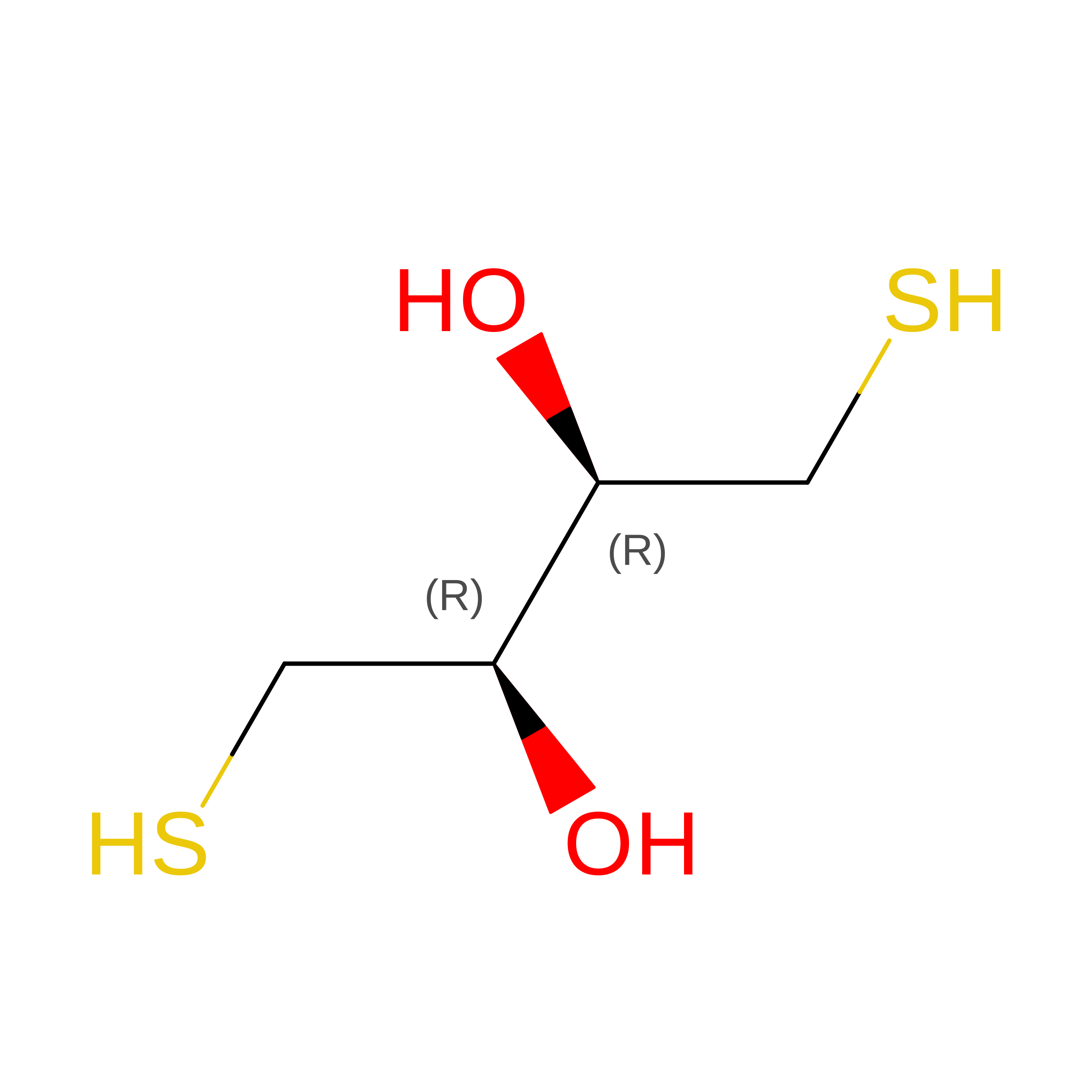
2,3-DIHYDROXY-1,4-DITHIOBUTANE
C4 H10 O2 S2
VHJLVAABSRFDPM-IMJSIDKUSA-N
GOL

Query on GOL


Download:Ideal Coordinates CCD File
J [auth A],
S [auth C],
V [auth D]
GLYCEROL
C3 H8 O3
PEDCQBHIVMGVHV-UHFFFAOYSA-N
CL

Query on CL


Download:Ideal Coordinates CCD File
G [auth A],
H [auth A],
Q [auth C]
CHLORIDE ION
Cl
VEXZGXHMUGYJMC-UHFFFAOYSA-M
NA

Query on NA


Download:Ideal Coordinates CCD File
E [auth A]
F [auth A]
K [auth B]
L [auth B]
N [auth C]
E [auth A],
F [auth A],
K [auth B],
L [auth B],
N [auth C],
O [auth C],
P [auth C],
T [auth D]
SODIUM ION
Na
FKNQFGJONOIPTF-UHFFFAOYSA-N

Experimental Data & Validation

Experimental Data

  • Method: X-RAY DIFFRACTION
  • Resolution: 1.47 Å
  • R-Value Free:  0.207 (Depositor), 0.212 (DCC) 
  • R-Value Work:  0.150 (Depositor), 0.153 (DCC) 
  • R-Value Observed: 0.150 (Depositor) 
Space Group: P 1
Unit Cell:
Length ( Å )Angle ( ˚ )
a = 55.3α = 92.16
b = 60.65β = 103.45
c = 98.71γ = 94.25
Software Package:
Software NamePurpose
REFMACrefinement
XDSdata reduction
XDSdata scaling
MOLREPphasing

Structure Validation

View Full Validation Report


Entry History 

Deposition Data

Revision History  (Full details and data files)

  • Version 1.0: 2006-10-17
    Type: Initial release
  • Version 1.1: 2008-04-30
    Changes: Version format compliance
  • Version 1.2: 2011-07-13
    Changes: Non-polymer description, Version format compliance
  • Version 1.3: 2016-05-18
    Changes: Database references
  • Version 1.4: 2023-10-25
    Changes: Data collection, Database references, Derived calculations, Refinement description