2A25 | pdb_00002a25

Crystal structure of Siah1 SBD bound to the peptide EKPAAVVAPITTG from SIP


Experimental Data Snapshot

  • Method: X-RAY DIFFRACTION
  • Resolution: 2.20 Å
  • R-Value Free: 
    0.252 (Depositor), 0.240 (DCC) 
  • R-Value Work: 
    0.213 (Depositor), 0.211 (DCC) 
  • R-Value Observed: 
    0.215 (Depositor) 

Starting Model: experimental
View more details

wwPDB Validation 3D Report Full Report

Validation slider image for 2A25

This is version 1.3 of the entry. See complete history

Literature

Structural Analysis of Siah1-Siah-interacting Protein Interactions and Insights into the Assembly of an E3 Ligase Multiprotein Complex

Santelli, E.Leone, M.Li, C.Fukushima, T.Preece, N.E.Olson, A.J.Ely, K.R.Reed, J.C.Pellecchia, M.Liddington, R.C.Matsuzawa, S.

(2005) J Biological Chem 280: 34278-34287

  • DOI: https://doi.org/10.1074/jbc.M506707200
  • Primary Citation Related Structures: 
    2A25, 2A26

  • PubMed Abstract: 

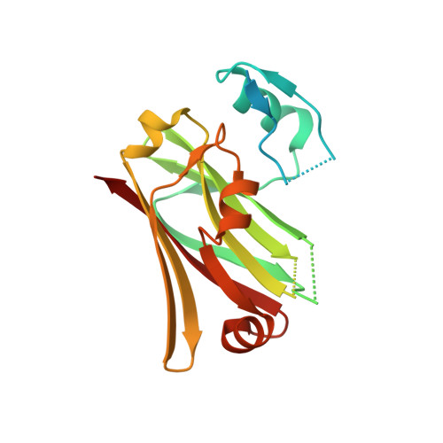
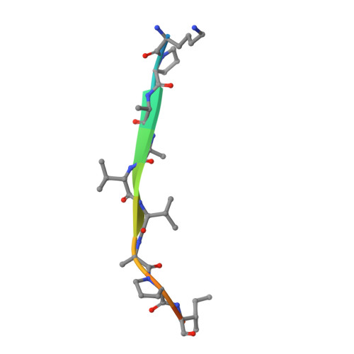
    Siah1 is the central component of a multiprotein E3 ubiquitin ligase complex that targets beta-catenin for destruction in response to p53 activation. The E3 complex comprises, in addition to Siah1, Siah-interacting protein (SIP), the adaptor protein Skp1, and the F-box protein Ebi. Here we show that SIP engages Siah1 by means of two elements, both of which are required for mediating beta-catenin destruction in cells. An N-terminal dimerization domain of SIP sits across the saddle-shaped upper surface of Siah1, with two extended legs packing against the sides of Siah1 by means of a consensus PXAXVXP motif that is common to a family of Siah-binding proteins. The C-terminal domain of SIP, which binds to Skp1, protrudes from the lower surface of Siah1, and we propose that this surface provides the scaffold for bringing substrate and the E2 enzyme into apposition in the functional complex.


  • Organizational Affiliation
    • The Burnham Institute, La Jolla, California 92037, USA.

Macromolecule Content 

  • Total Structure Weight: 23.01 kDa 
  • Atom Count: 1,320 
  • Modeled Residue Count: 155 
  • Deposited Residue Count: 206 
  • Unique protein chains: 2

Macromolecules

Find similar proteins by:|  3D Structure
Entity ID: 1
MoleculeChains  Sequence LengthOrganismDetailsImage
Ubiquitin ligase SIAH1193Homo sapiensMutation(s): 0 
Gene Names: SIAH1HUMSIAH
EC: 6.3.2 (PDB Primary Data), 2.3.2.27 (UniProt)
UniProt & NIH Common Fund Data Resources
Find proteins for Q8IUQ4 (Homo sapiens)
Explore Q8IUQ4 
Go to UniProtKB:  Q8IUQ4
PHAROS:  Q8IUQ4
GTEx:  ENSG00000196470 
Entity Groups
Sequence Clusters30% Identity50% Identity70% Identity90% Identity95% Identity100% Identity
UniProt GroupQ8IUQ4
Sequence Annotations
Expand
Reference Sequence
Find similar proteins by:  Sequence   |   3D Structure  
Entity ID: 2
MoleculeChains  Sequence LengthOrganismDetailsImage
Calcyclin-binding protein peptide13N/AMutation(s): 0 
UniProt & NIH Common Fund Data Resources
Find proteins for Q9HB71 (Homo sapiens)
Explore Q9HB71 
Go to UniProtKB:  Q9HB71
PHAROS:  Q9HB71
GTEx:  ENSG00000116161 
Entity Groups
Sequence Clusters30% Identity50% Identity70% Identity90% Identity95% Identity100% Identity
UniProt GroupQ9HB71
Sequence Annotations
Expand
Reference Sequence

Small Molecules

Ligands 1 Unique
IDChains Name / Formula / InChI Key2D Diagram3D Interactions
ZN

Query on ZN


Download:Ideal Coordinates CCD File
C [auth A]ZINC ION
Zn
PTFCDOFLOPIGGS-UHFFFAOYSA-N

Experimental Data & Validation

Experimental Data

  • Method: X-RAY DIFFRACTION
  • Resolution: 2.20 Å
  • R-Value Free:  0.252 (Depositor), 0.240 (DCC) 
  • R-Value Work:  0.213 (Depositor), 0.211 (DCC) 
  • R-Value Observed: 0.215 (Depositor) 
Space Group: C 2 2 21
Unit Cell:
Length ( Å )Angle ( ˚ )
a = 47.635α = 90
b = 107.761β = 90
c = 78.414γ = 90
Software Package:
Software NamePurpose
REFMACrefinement
DENZOdata reduction
SCALEPACKdata scaling
MOLREPphasing

Structure Validation

View Full Validation Report


Entry History 

Deposition Data

Revision History  (Full details and data files)

  • Version 1.0: 2005-08-09
    Type: Initial release
  • Version 1.1: 2008-04-30
    Changes: Version format compliance
  • Version 1.2: 2011-07-13
    Changes: Advisory, Version format compliance
  • Version 1.3: 2023-08-23
    Changes: Data collection, Database references, Derived calculations, Refinement description