2A15 | pdb_00002a15

X-ray Crystal Structure of RV0760 from Mycobacterium Tuberculosis at 1.68 Angstrom Resolution


Experimental Data Snapshot

  • Method: X-RAY DIFFRACTION
  • Resolution: 1.68 Å
  • R-Value Free: 
    0.207 (Depositor), 0.179 (DCC) 
  • R-Value Work: 
    0.176 (Depositor), 0.178 (DCC) 
  • R-Value Observed: 
    0.178 (Depositor) 

Starting Model: experimental
View more details

wwPDB Validation 3D Report Full Report

Validation slider image for 2A15

This is version 1.6 of the entry. See complete history

Literature

Crystal structure of Mycobacterium tuberculosis Rv0760c at 1.50 A resolution, a structural homolog of Delta(5)-3-ketosteroid isomerase

Cherney, M.M.Garen, C.R.James, M.N.G.

(2008) Biochim Biophys Acta 1784: 1625-1632

  • DOI: https://doi.org/10.1016/j.bbapap.2008.05.012
  • Primary Citation Related Structures: 
    2A15, 2Z76, 2Z77, 2Z7A

  • PubMed Abstract: 

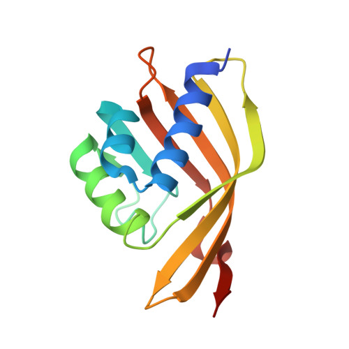
    We have determined the X-ray crystal structure of the Mycobacterium tuberculosis (Mtb) gene product encoded by the open reading frame Rv0760c at 1.50 A resolution by single-wavelength anomalous dispersion (SAD) phasing of diffraction data from crystals of the selenomethionine-substituted protein. Refinement against diffraction data from the native protein resulted in R(work)=19.5% and R(free)=21.4%. The X-ray crystal structure shows that the homodimeric Rv0760c polypeptide has an alpha + beta conical barrel fold placing it among many structural neighbors of the nuclear transport factor 2 family (NTF2). This family is highly conserved in terms of structure; however the substrates and individual protein functions are diverse. The structures of native Rv0760c in several different crystal forms and Rv0760c bound to 17beta-estradiol 17-hemisuccinate (EH) have also been solved and analyzed.


  • Organizational Affiliation
    • Group in Protein Structure and Function, Department of Biochemistry, University of Alberta, Edmonton, Alberta, Canada T6G 2H7.

Macromolecule Content 

  • Total Structure Weight: 15.38 kDa 
  • Atom Count: 1,258 
  • Modeled Residue Count: 133 
  • Deposited Residue Count: 139 
  • Unique protein chains: 1

Macromolecules

Find similar proteins by:|  3D Structure
Entity ID: 1
MoleculeChains  Sequence LengthOrganismDetailsImage
HYPOTHETICAL PROTEIN Rv0760c139Mycobacterium tuberculosisMutation(s): 0 
Gene Names: Rv0760
UniProt
Find proteins for P71817 (Mycobacterium tuberculosis (strain CDC 1551 / Oshkosh))
Explore P71817 
Go to UniProtKB:  P71817
Entity Groups
Sequence Clusters30% Identity50% Identity70% Identity90% Identity95% Identity100% Identity
UniProt GroupP71817
Sequence Annotations
Expand
Reference Sequence

Small Molecules

Ligands 1 Unique
IDChains Name / Formula / InChI Key2D Diagram3D Interactions
NCA

Query on NCA


Download:Ideal Coordinates CCD File
B [auth A]NICOTINAMIDE
C6 H6 N2 O
DFPAKSUCGFBDDF-UHFFFAOYSA-N

Experimental Data & Validation

Experimental Data

  • Method: X-RAY DIFFRACTION
  • Resolution: 1.68 Å
  • R-Value Free:  0.207 (Depositor), 0.179 (DCC) 
  • R-Value Work:  0.176 (Depositor), 0.178 (DCC) 
  • R-Value Observed: 0.178 (Depositor) 
Space Group: P 32 1 2
Unit Cell:
Length ( Å )Angle ( ˚ )
a = 55.6α = 90
b = 55.6β = 90
c = 104.74γ = 120
Software Package:
Software NamePurpose
REFMACrefinement
SCALEPACKdata scaling
MOLREPphasing
ELVESrefinement
ELVESdata reduction

Structure Validation

View Full Validation Report


Entry History 

Deposition Data

Revision History  (Full details and data files)

  • Version 1.0: 2005-10-11
    Type: Initial release
  • Version 1.1: 2008-04-30
    Changes: Version format compliance
  • Version 1.2: 2011-07-13
    Changes: Version format compliance
  • Version 1.3: 2017-10-11
    Changes: Refinement description
  • Version 1.4: 2019-07-24
    Changes: Data collection, Refinement description
  • Version 1.5: 2024-02-14
    Changes: Data collection, Database references, Derived calculations
  • Version 1.6: 2024-04-03
    Changes: Refinement description