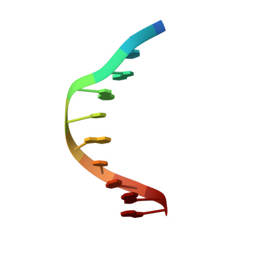
Crystal structure of a covalent DNA-drug adduct: anthramycin bound to C-C-A-A-C-G-T-T-G-G and a molecular explanation of specificity.
Kopka, M.L., Goodsell, D.S., Baikalov, I., Grzeskowiak, K., Cascio, D., Dickerson, R.E.(1994) Biochemistry 33: 13593-13610
- PubMed: 7947769 Search on PubMed
- DOI: https://doi.org/10.1021/bi00250a011
- Primary Citation Related Structures:
274D - PubMed Abstract:
A 2.3-A X-ray crystal structure analysis has been carried out on the antitumor drug anthramycin, covalently bound to a ten base pair DNA double helix of sequence C-C-A-A-C-G-T-T-G-G. One drug molecule sits within the minor groove at each end of the helix, covalently bound through its C11 position to the N2 amine of the penultimate guanine of the chain. The stereochemical conformation is C11S, C11aS. The natural twist of the anthramycin molecule in the C11aS conformation matches the twist of the minor groove as it winds along the helix; a C11aR drug would only fit into a left-handed helix. The C11S attachment is roughly equatorial to the overall plane of the molecule, whereas a C11R attachment would be axial and would obstruct the fitting of the drug into the groove. The six-membered ring of anthramycin points toward the 3' end of the chain to which it is covalently attached or toward the end of the helix. The acrylamide tail attached to the five-membered ring extends back along the minor groove toward the center of the helix, binding in a manner reminiscent of netropsin or distamycin. The drug-DNA complex is stabilized by hydrogen bonds from C9-OH, N10, and the end of the acrylamide tail to base pair edges on the floor of the minor groove. The origin of anthramycin specificity for three successive purines arises not from specific hydrogen bonds but from the low twist angles adopted by purine-purine steps in a B-DNA helix. Binding of anthramycin induces a low twist at T-G in the T-G-G sequence of this DNA-drug complex, by comparison with the structure of the free DNA. The origin of anthramycin's preference for adenines flanking the alkylated guanine arises from a netropsin-like fitting of the acrylamide tail into the minor groove.
- Department of Chemistry and Biochemistry, University of California, Los Angeles 90024.
Organizational Affiliation: