1Z70

1.15A resolution structure of the formylglycine generating enzyme FGE


Experimental Data Snapshot

  • Method: X-RAY DIFFRACTION
  • Resolution: 1.15 Å
  • R-Value Free: 0.170 
  • R-Value Work: 0.138 
  • R-Value Observed: 0.139 

wwPDB Validation   3D Report Full Report


Ligand Structure Quality Assessment 


This is version 2.2 of the entry. See complete history


Literature

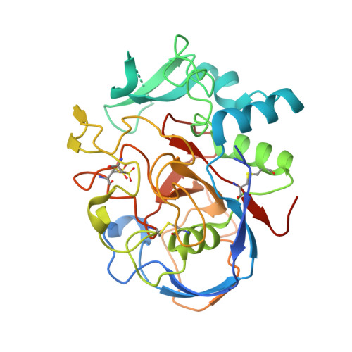
De novo calcium/sulfur SAD phasing of the human formylglycine-generating enzyme using in-house data.

Roeser, D.Dickmanns, A.Gasow, K.Rudolph, M.G.

(2005) Acta Crystallogr D Biol Crystallogr 61: 1057-1066

  • DOI: https://doi.org/10.1107/S0907444905013831
  • Primary Citation of Related Structures:  
    1Z70

  • PubMed Abstract: 

    Sulfatases are a family of enzymes essential for the degradation of sulfate esters. Formylglycine is the key catalytic residue in the active site of sulfatases and is generated from a cysteine residue by FGE, the formylglycine-generating enzyme. Inactivity of FGE owing to inherited mutations in the FGE gene results in multiple sulfatase deficiency (MSD), which leads to early death in infants. Human FGE was crystallized in the presence of traces of the protease elastase, which was absolutely essential for crystal growth, and the structure of FGE was determined by molecular replacement. Before this model was completed, the FGE structure was re-determined by SAD phasing using in-house data based on the anomalous signal of two calcium ions bound to the native enzyme and intrinsic S atoms. A 14-atom substructure was determined at 1.8 A resolution by SHELXD; SHELXE was used for density modification and phase extension to 1.54 A resolution. Automated model building with ARP/wARP and refinement with REFMAC5 yielded a virtually complete model without manual intervention. The minimal data requirements for successful phasing and the relative contributions of the Ca and S atoms are discussed and compared with the related FGE paralogue, pFGE. This work emphasizes the usefulness of de novo phasing using weak anomalous scatterers and in-house data.


  • Organizational Affiliation

    Department of Molecular Structural Biology, University of Göttingen, D-37077 Göttingen, Germany.


Macromolecules
Find similar proteins by:  (by identity cutoff)  |  3D Structure
Entity ID: 1
MoleculeChains Sequence LengthOrganismDetailsImage
C-alpha-formyglycine-generating enzymeA [auth X]311Homo sapiensMutation(s): 2 
UniProt & NIH Common Fund Data Resources
Find proteins for Q8NBK3 (Homo sapiens)
Explore Q8NBK3 
Go to UniProtKB:  Q8NBK3
PHAROS:  Q8NBK3
GTEx:  ENSG00000144455 
Entity Groups  
Sequence Clusters30% Identity50% Identity70% Identity90% Identity95% Identity100% Identity
UniProt GroupQ8NBK3
Sequence Annotations
Expand
  • Reference Sequence
Oligosaccharides

Help

Entity ID: 2
MoleculeChains Length2D Diagram Glycosylation3D Interactions
2-acetamido-2-deoxy-beta-D-glucopyranose-(1-4)-2-acetamido-2-deoxy-beta-D-glucopyranoseB [auth A]2N-Glycosylation
Glycosylation Resources
GlyTouCan:  G42666HT
GlyCosmos:  G42666HT
GlyGen:  G42666HT
Small Molecules
Modified Residues  1 Unique
IDChains TypeFormula2D DiagramParent
OCS
Query on OCS
A [auth X]L-PEPTIDE LINKINGC3 H7 N O5 SCYS
Experimental Data & Validation

Experimental Data

  • Method: X-RAY DIFFRACTION
  • Resolution: 1.15 Å
  • R-Value Free: 0.170 
  • R-Value Work: 0.138 
  • R-Value Observed: 0.139 
  • Space Group: P 21 21 2
Unit Cell:
Length ( Å )Angle ( ˚ )
a = 62.295α = 90
b = 109.765β = 90
c = 43.511γ = 90
Software Package:
Software NamePurpose
REFMACrefinement
HKL-2000data reduction
SCALEPACKdata scaling

Structure Validation

View Full Validation Report


Ligand Structure Quality Assessment 


Entry History 

Deposition Data

Revision History  (Full details and data files)

  • Version 1.0: 2005-07-22
    Type: Initial release
  • Version 1.1: 2008-04-30
    Changes: Version format compliance
  • Version 1.2: 2011-07-13
    Changes: Non-polymer description, Version format compliance
  • Version 2.0: 2020-07-29
    Type: Remediation
    Reason: Carbohydrate remediation
    Changes: Atomic model, Data collection, Database references, Derived calculations, Structure summary
  • Version 2.1: 2023-08-23
    Changes: Data collection, Database references, Refinement description, Structure summary
  • Version 2.2: 2023-11-15
    Changes: Data collection