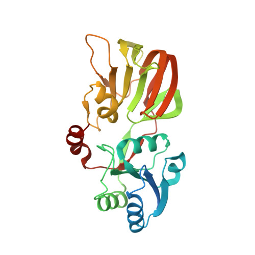
1Z0S

Crystal structure of an NAD kinase from Archaeoglobus fulgidus in complex with ATP


Experimental Data Snapshot

  • Method: X-RAY DIFFRACTION
  • Resolution: 1.70 Å
  • R-Value Free: 0.231 
  • R-Value Work: 0.200 
  • R-Value Observed: 0.200 

wwPDB Validation   3D Report Full Report


Ligand Structure Quality Assessment 


This is version 1.3 of the entry. See complete history


Literature

Crystal Structures of an NAD Kinase from Archaeoglobus fulgidus in Complex with ATP, NAD, or NADP

Liu, J.Lou, Y.Yokota, H.Adams, P.D.Kim, R.Kim, S.H.

(2005) J Mol Biol 354: 289-303

  • DOI: https://doi.org/10.1016/j.jmb.2005.09.026
  • Primary Citation of Related Structures:  
    1SUW, 1Z0S, 1Z0U, 1Z0Z

  • PubMed Abstract: 

    NAD kinase is a ubiquitous enzyme that catalyzes the phosphorylation of NAD to NADP using ATP or inorganic polyphosphate (poly(P)) as phosphate donor, and is regarded as the only enzyme responsible for the synthesis of NADP. We present here the crystal structures of an NAD kinase from the archaeal organism Archaeoglobus fulgidus in complex with its phosphate donor ATP at 1.7 A resolution, with its substrate NAD at 3.05 A resolution, and with the product NADP in two different crystal forms at 2.45 A and 2.0 A resolution, respectively. In the ATP bound structure, the AMP portion of the ATP molecule is found to use the same binding site as the nicotinamide ribose portion of NAD/NADP in the NAD/NADP bound structures. A magnesium ion is found to be coordinated to the phosphate tail of ATP as well as to a pyrophosphate group. The conserved GGDG loop forms hydrogen bonds with the pyrophosphate group in the ATP-bound structure and the 2' phosphate group of the NADP in the NADP-bound structures. A possible phosphate transfer mechanism is proposed on the basis of the structures presented.


  • Organizational Affiliation

    Berkeley Structural Genomics Center, Lawrence Berkeley National Laboratory, Physical Bioscience Division, Berkeley, CA 94720-5230, USA.


Macromolecules
Find similar proteins by:  (by identity cutoff)  |  3D Structure
Entity ID: 1
MoleculeChains Sequence LengthOrganismDetailsImage
Probable inorganic polyphosphate/ATP-NAD kinase
A, B, C, D
278Archaeoglobus fulgidusMutation(s): 0 
Gene Names: ppnK
EC: 2.7.1.23
UniProt
Find proteins for O30297 (Archaeoglobus fulgidus (strain ATCC 49558 / DSM 4304 / JCM 9628 / NBRC 100126 / VC-16))
Explore O30297 
Go to UniProtKB:  O30297
Entity Groups  
Sequence Clusters30% Identity50% Identity70% Identity90% Identity95% Identity100% Identity
UniProt GroupO30297
Sequence Annotations
Expand
  • Reference Sequence
Small Molecules
Ligands 3 Unique
IDChains Name / Formula / InChI Key2D Diagram3D Interactions
ATP
Query on ATP

Download Ideal Coordinates CCD File 
F [auth A],
I [auth B],
L [auth C],
O [auth D]
ADENOSINE-5'-TRIPHOSPHATE
C10 H16 N5 O13 P3
ZKHQWZAMYRWXGA-KQYNXXCUSA-N
POP
Query on POP

Download Ideal Coordinates CCD File 
G [auth A],
J [auth B],
M [auth C],
P [auth D]
PYROPHOSPHATE 2-
H2 O7 P2
XPPKVPWEQAFLFU-UHFFFAOYSA-L
MG
Query on MG

Download Ideal Coordinates CCD File 
E [auth A],
H [auth B],
K [auth C],
N [auth D]
MAGNESIUM ION
Mg
JLVVSXFLKOJNIY-UHFFFAOYSA-N
Experimental Data & Validation

Experimental Data

  • Method: X-RAY DIFFRACTION
  • Resolution: 1.70 Å
  • R-Value Free: 0.231 
  • R-Value Work: 0.200 
  • R-Value Observed: 0.200 
  • Space Group: P 1
Unit Cell:
Length ( Å )Angle ( ˚ )
a = 55.252α = 106.35
b = 69.731β = 111.32
c = 76.287γ = 101.3
Software Package:
Software NamePurpose
HKL-2000data collection
SCALEPACKdata scaling
AMoREphasing
REFMACrefinement
HKL-2000data reduction

Structure Validation

View Full Validation Report


Ligand Structure Quality Assessment 


Entry History 

Deposition Data

Revision History  (Full details and data files)

  • Version 1.0: 2005-04-19
    Type: Initial release
  • Version 1.1: 2008-04-30
    Changes: Version format compliance
  • Version 1.2: 2011-07-13
    Changes: Version format compliance
  • Version 1.3: 2024-02-14
    Changes: Data collection, Database references, Derived calculations