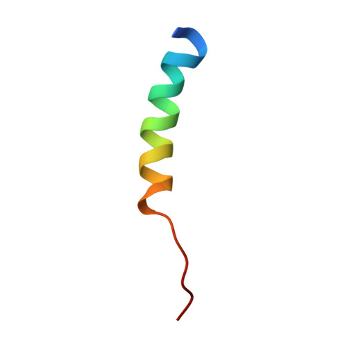
The N-terminal 26-residue fragment of human programmed cell death 5 protein can form a stable alpha-helix having unique electrostatic potential character.
Liu, D., Yao, H., Chen, Y., Feng, Y., Chen, Y., Wang, J.(2005) Biochem J 392: 47-54
- PubMed: 16083422 Search on PubMedSearch on PubMed Central
- DOI: https://doi.org/10.1042/BJ20050688
- Primary Citation Related Structures:
1YYB - PubMed Abstract:
PDCD5-(1-26) is a N-terminal 26-residue fragment of human PDCD5 (programmed cell death 5) protein. PDCD5 is an important novel protein that regulates both apoptotic and non-apoptotic programmed cell death. The conformation of PDCD5 protein is a stable helical core consisting of a triple-helix bundle and two dissociated terminal regions. The N-terminal region is ordered and contains abundant secondary structure. Overexpression and purification of the N-terminal 26-residure fragment, PDCD5-(1-26), was performed in this study to better understand its tertiary structure. The spectroscopic studies using CD and hetero- and homo-nuclear NMR methods determine a stable alpha-helix formed by Asp3-Ala19 of PDCD5-(1-26). The N-terminal residues Asp3-Ala19 of PDCD5 were then affirmed to have the capacity to form a stable alpha-helix independently of the core of the protein. Analysis of the helical peptide of PDCD5-(1-26) indicates that the surface of this well-formed alpha-helix has a unique electrostatic potential character. This may provide an environment for the N-terminal alpha-helix of PDCD5 to serve as an independent functional entity of the protein. The apoptosis activity assay shows that the deletion of the N-terminal alpha-helix of PDCD5 significantly attenuates the apoptosis-promoting effects on HL-60 cells induced by serum withdrawal.
- National Laboratory of Biomacromolecules, Center for Molecular Biology, Institute of Biophysics, Chinese Academy of Sciences, 15 Datun Road, Beijing 100101, China.
Organizational Affiliation: