Structure, epitope mapping, and docking simulation of a gibberellin mimic peptide as a peptidyl mimotope for a hydrophobic ligand.
Murata, T., Hemmi, H., Nakamura, S., Shimizu, K., Suzuki, Y., Yamaguchi, I.(2005) FEBS J 272: 4938-4948
- PubMed: 16176267 Search on PubMed
- DOI: https://doi.org/10.1111/j.1742-4658.2005.04902.x
- Primary Citation Related Structures:
1YT6 - PubMed Abstract:
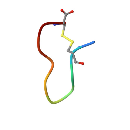
Using NMR spectroscopy and simulated annealing calculations, we determined the solution structure of the disulfide-linked cyclized decapeptide ACLPWSDGPC (SD), which is bound to an anti-(gibberellin A(4)) mAb 4-B8(8)/E9 and was found to be the first peptidyl mimotope for a hydrophobic ligand. The resulting structure of the peptide showed a beta-turn-like conformation in residues three to seven and the region converges well (average rmsd 0.54 A). The binding activity and the epitopes of the peptide to the antibody were assessed using saturation transfer difference (STD)-NMR experiments. We also conducted docking simulations between the peptide and the mAb to determine how the peptide is bound to the mAb. Resonances around the beta-turn-like conformation of peptide SD (residues 3-5) showed strong STD enhancement, which agreed well with results from docking simulation between peptide SD and the mAb. Together with the commonality of amino acid residues of the mAb involved in interactions with gibberellin A(4) (GA(4)) and peptide SD, we concluded that peptide SD is bound to the antigen-binding site of mAb 4-B8(8)/E9 as a GA(4) mimic, confirming evidence for the existence of peptide mimics even for hydrophobic ligands.
- National Food Research Institute, Kannondai, Tsukuba, Japan.
Organizational Affiliation: