1YRO | pdb_00001yro

Crystal structure of beta14,-galactosyltransferase mutant ARG228Lys in complex with alpha-lactalbumin in the presence of UDP-galactose and Mn


Experimental Data Snapshot

  • Method: X-RAY DIFFRACTION
  • Resolution: 1.90 Å
  • R-Value Free: 
    0.225 (Depositor), 0.221 (DCC) 
  • R-Value Work: 
    0.189 (Depositor), 0.185 (DCC) 
  • R-Value Observed: 
    0.189 (Depositor) 

Starting Model: experimental
View more details

wwPDB Validation 3D Report Full Report

Validation slider image for 1YRO

Ligand Structure Quality Assessment 


This is version 1.6 of the entry. See complete history

Literature

Mutation of Arginine 228 to Lysine Enhances the Glucosyltransferase Activity of Bovine beta-1,4-Galactosyltransferase I

Ramakrishnan, B.Boeggeman, E.Qasba, P.K.

(2005) Biochemistry 44: 3202-3210

  • DOI: https://doi.org/10.1021/bi0479454
  • Primary Citation Related Structures: 
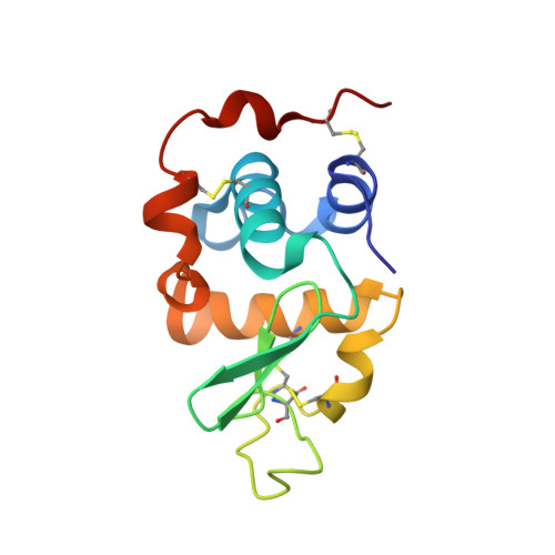
    1YRO

  • PubMed Abstract: 

    Beta-1,4-galactosyltransferase I (beta4Gal-T1) normally transfers Gal from UDP-Gal to GlcNAc in the presence of Mn(2+) ion (Gal-T activity) and also transfers Glc from UDP-Glc to GlcNAc (Glc-T activity), albeit at only 0.3% efficiency. In addition, alpha-lactalbumin (LA) enhances this Glc-T activity more than 25 times. Comparison of the crystal structures of UDP-Gal- and UDP-Glc-bound beta4Gal-T1 reveals that the O4 hydroxyl group in both Gal and Glc moieties forms a hydrogen bond with the side chain carboxylate group of Glu317. The orientation of the O4 hydroxyl of glucose causes a steric hindrance to the side chain carboxylate group of Glu317, accounting for the enzyme's low Glc-T activity. In this study, we show that mutation of Arg228, a residue in the vicinity of Glu317, to lysine (R228K-Gal-T1) results in a 15-fold higher Glc-T activity, which is further enhanced by LA to nearly 25% of the Gal-T activity of the wild type. The kinetic parameters indicate that the main effect of the mutation of Arg228 to lysine is on the k(cat) of Glc-T, which increases 3-4-fold, both in the absence and in the presence of LA; simultaneously, the k(cat) for the Gal-T reaction is reduced 30-fold. The crystal structure of R228K-Gal-T1 complexed with LA, UDP-Gal, and Mn(2+) determined at 1.9 A resolution shows that the Asp318 side chain exhibits a minor alternate conformation, compared to that in the wild type. This alternate conformation now causes a steric hindrance to the O4 hydroxyl group of the Gal moiety of UDP-Gal, probably causing the dissociation of UDP-Gal and the reduced k(cat) of the Gal-T reaction.


  • Organizational Affiliation
    • Structural Glycobiology Section, Laboratory of Experimental and Computational Biology, Center for Cancer Research, National Cancer Institute-Frederick, Frederick, Maryland 21702-1201, USA.

Macromolecule Content 

  • Total Structure Weight: 96.39 kDa 
  • Atom Count: 7,388 
  • Modeled Residue Count: 790 
  • Deposited Residue Count: 818 
  • Unique protein chains: 2

Macromolecules

Find similar proteins by:|  3D Structure
Entity ID: 1
MoleculeChains  Sequence LengthOrganismDetailsImage
ALPHA-LACTALBUMIN
A, C
123Mus musculusMutation(s): 0 
Gene Names: alpha-lactalbumin
UniProt
Find proteins for P29752 (Mus musculus)
Explore P29752 
Go to UniProtKB:  P29752
Entity Groups
Sequence Clusters30% Identity50% Identity70% Identity90% Identity95% Identity100% Identity
UniProt GroupP29752
Sequence Annotations
Expand
Reference Sequence
Find similar proteins by:|  3D Structure
Entity ID: 2
MoleculeChains  Sequence LengthOrganismDetailsImage
BETA-1,4-GALACTOSYLTRANSFERASE
B, D
286Bos taurusMutation(s): 2 
Gene Names: beta14-galactosyltransferase
EC: 2.4.1.22 (PDB Primary Data), 2.4.1.90 (PDB Primary Data), 2.4.1.38 (PDB Primary Data), 2.4.1 (UniProt), 2.4.1.275 (UniProt)
UniProt
Find proteins for P08037 (Bos taurus)
Explore P08037 
Go to UniProtKB:  P08037
Entity Groups
Sequence Clusters30% Identity50% Identity70% Identity90% Identity95% Identity100% Identity
UniProt GroupP08037
Sequence Annotations
Expand
Reference Sequence

Small Molecules

Ligands 6 Unique
IDChains Name / Formula / InChI Key2D Diagram3D Interactions
GDU

Query on GDU


Download:Ideal Coordinates CCD File
H [auth B],
M [auth D]
GALACTOSE-URIDINE-5'-DIPHOSPHATE
C15 H24 N2 O17 P2
HSCJRCZFDFQWRP-ABVWGUQPSA-N
UDP

Query on UDP


Download:Ideal Coordinates CCD File
J [auth B],
O [auth D]
URIDINE-5'-DIPHOSPHATE
C9 H14 N2 O12 P2
XCCTYIAWTASOJW-XVFCMESISA-N
MES

Query on MES


Download:Ideal Coordinates CCD File
F [auth A]2-(N-MORPHOLINO)-ETHANESULFONIC ACID
C6 H13 N O4 S
SXGZJKUKBWWHRA-UHFFFAOYSA-N
PG4

Query on PG4


Download:Ideal Coordinates CCD File
G [auth A],
L [auth C]
TETRAETHYLENE GLYCOL
C8 H18 O5
UWHCKJMYHZGTIT-UHFFFAOYSA-N
MN

Query on MN


Download:Ideal Coordinates CCD File
I [auth B],
N [auth D]
MANGANESE (II) ION
Mn
WAEMQWOKJMHJLA-UHFFFAOYSA-N
CA

Query on CA


Download:Ideal Coordinates CCD File
E [auth A],
K [auth C]
CALCIUM ION
Ca
BHPQYMZQTOCNFJ-UHFFFAOYSA-N

Experimental Data & Validation

Experimental Data

  • Method: X-RAY DIFFRACTION
  • Resolution: 1.90 Å
  • R-Value Free:  0.225 (Depositor), 0.221 (DCC) 
  • R-Value Work:  0.189 (Depositor), 0.185 (DCC) 
  • R-Value Observed: 0.189 (Depositor) 
Space Group: P 1 21 1
Unit Cell:
Length ( Å )Angle ( ˚ )
a = 57.334α = 90
b = 94.087β = 101.64
c = 99.705γ = 90
Software Package:
Software NamePurpose
HKL-2000data collection
SCALEPACKdata scaling
AMoREphasing
CNSrefinement
HKL-2000data reduction

Structure Validation

View Full Validation Report


Ligand Structure Quality Assessment 


Entry History 

Deposition Data

Revision History  (Full details and data files)

  • Version 1.0: 2005-03-22
    Type: Initial release
  • Version 1.1: 2007-10-16
    Changes: Version format compliance
  • Version 1.2: 2011-07-13
    Changes: Version format compliance
  • Version 1.3: 2017-10-11
    Changes: Data collection
  • Version 1.4: 2021-10-20
    Changes: Database references, Derived calculations
  • Version 1.5: 2023-08-23
    Changes: Data collection, Refinement description
  • Version 1.6: 2024-11-06
    Changes: Structure summary