1Y8O

Crystal structure of the PDK3-L2 complex


Experimental Data Snapshot

  • Method: X-RAY DIFFRACTION
  • Resolution: 2.48 Å
  • R-Value Free: 0.234 
  • R-Value Work: 0.210 
  • R-Value Observed: 0.211 

wwPDB Validation   3D Report Full Report


This is version 1.3 of the entry. See complete history


Literature

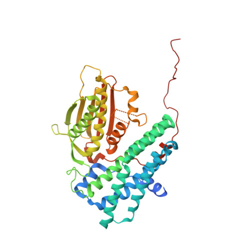
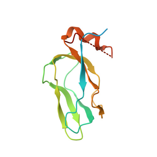
Crystal structure of pyruvate dehydrogenase kinase 3 bound to lipoyl domain 2 of human pyruvate dehydrogenase complex.

Kato, M.Chuang, J.L.Tso, S.C.Wynn, R.M.Chuang, D.T.

(2005) EMBO J 24: 1763-1774

  • DOI: https://doi.org/10.1038/sj.emboj.7600663
  • Primary Citation of Related Structures:  
    1Y8N, 1Y8O, 1Y8P

  • PubMed Abstract: 
  • The human pyruvate dehydrogenase complex (PDC) is regulated by reversible phosphorylation by four isoforms of pyruvate dehydrogenase kinase (PDK). PDKs phosphorylate serine residues in the dehydrogenase (E1p) component of PDC, but their amino-acid sequences are unrelated to eukaryotic Ser/Thr/Tyr protein kinases ...

    The human pyruvate dehydrogenase complex (PDC) is regulated by reversible phosphorylation by four isoforms of pyruvate dehydrogenase kinase (PDK). PDKs phosphorylate serine residues in the dehydrogenase (E1p) component of PDC, but their amino-acid sequences are unrelated to eukaryotic Ser/Thr/Tyr protein kinases. PDK3 binds to the inner lipoyl domains (L2) from the 60-meric transacetylase (E2p) core of PDC, with concomitant stimulated kinase activity. Here, we present crystal structures of the PDK3-L2 complex with and without bound ADP or ATP. These structures disclose that the C-terminal tail from one subunit of PDK3 dimer constitutes an integral part of the lipoyl-binding pocket in the N-terminal domain of the opposing subunit. The two swapped C-terminal tails promote conformational changes in active-site clefts of both PDK3 subunits, resulting in largely disordered ATP lids in the ADP-bound form. Our structural and biochemical data suggest that L2 binding stimulates PDK3 activity by disrupting the ATP lid, which otherwise traps ADP, to remove product inhibition exerted by this nucleotide. We hypothesize that this allosteric mechanism accounts, in part, for E2p-augmented PDK3 activity.


    Organizational Affiliation

    Department of Internal Medicine, University of Texas Southwestern Medical Center, 5323 Harry Hines Boulevard, Dallas, TX 75390, USA.


Macromolecules
Find similar proteins by:  (by identity cutoff)  |  3D Structure
Entity ID: 1
MoleculeChainsSequence LengthOrganismDetailsImage
[Pyruvate dehydrogenase [lipoamide]] kinase isozyme 3419Homo sapiensMutation(s): 0 
Gene Names: PDK3PDHK3
EC: 2.7.1.99 (PDB Primary Data), 2.7.11.2 (UniProt)
UniProt & NIH Common Fund Data Resources
Find proteins for Q15120 (Homo sapiens)
Explore Q15120 
Go to UniProtKB:  Q15120
PHAROS:  Q15120
Entity Groups  
Sequence Clusters30% Identity50% Identity70% Identity90% Identity95% Identity100% Identity
UniProt GroupQ15120
Protein Feature View
Expand
  • Reference Sequence
Find similar proteins by:  (by identity cutoff)  |  3D Structure
Entity ID: 2
MoleculeChainsSequence LengthOrganismDetailsImage
Dihydrolipoyllysine-residue acetyltransferase component of pyruvate dehydrogenase complex128Homo sapiensMutation(s): 0 
Gene Names: DLATDLTA
EC: 2.3.1.12
UniProt & NIH Common Fund Data Resources
Find proteins for P10515 (Homo sapiens)
Explore P10515 
Go to UniProtKB:  P10515
PHAROS:  P10515
Entity Groups  
Sequence Clusters30% Identity50% Identity70% Identity90% Identity95% Identity100% Identity
UniProt GroupP10515
Protein Feature View
Expand
  • Reference Sequence
Small Molecules
Ligands 4 Unique
IDChainsName / Formula / InChI Key2D Diagram3D Interactions
ADP
Query on ADP

Download Ideal Coordinates CCD File 
F [auth A]ADENOSINE-5'-DIPHOSPHATE
C10 H15 N5 O10 P2
XTWYTFMLZFPYCI-KQYNXXCUSA-N
 Ligand Interaction
RED
Query on RED

Download Ideal Coordinates CCD File 
G [auth B]DIHYDROLIPOIC ACID
C8 H16 O2 S2
IZFHEQBZOYJLPK-SSDOTTSWSA-N
 Ligand Interaction
K
Query on K

Download Ideal Coordinates CCD File 
D [auth A],
E [auth A]
POTASSIUM ION
K
NPYPAHLBTDXSSS-UHFFFAOYSA-N
 Ligand Interaction
MG
Query on MG

Download Ideal Coordinates CCD File 
C [auth A]MAGNESIUM ION
Mg
JLVVSXFLKOJNIY-UHFFFAOYSA-N
 Ligand Interaction
Binding Affinity Annotations 
IDSourceBinding Affinity
ADP PDBBind:  1Y8O Kd: 1300 (nM) from 1 assay(s)
Binding MOAD:  1Y8O Kd: 1300 (nM) from 1 assay(s)
Experimental Data & Validation

Experimental Data

  • Method: X-RAY DIFFRACTION
  • Resolution: 2.48 Å
  • R-Value Free: 0.234 
  • R-Value Work: 0.210 
  • R-Value Observed: 0.211 
  • Space Group: P 65 2 2
Unit Cell:
Length ( Å )Angle ( ˚ )
a = 120.752α = 90
b = 120.752β = 90
c = 239.153γ = 120
Software Package:
Software NamePurpose
REFMACrefinement
HKL-2000data reduction
SCALEPACKdata scaling
AMoREphasing

Structure Validation

View Full Validation Report


Entry History 

Deposition Data

Revision History  (Full details and data files)

  • Version 1.0: 2005-05-24
    Type: Initial release
  • Version 1.1: 2008-04-30
    Changes: Version format compliance
  • Version 1.2: 2011-07-13
    Changes: Derived calculations, Version format compliance
  • Version 1.3: 2011-10-05
    Changes: Non-polymer description