Role of the divalent metal ion in sugar binding, ring opening, and isomerization by D-xylose isomerase: replacement of a catalytic metal by an amino acid.
Allen, K.N., Lavie, A., Glasfeld, A., Tanada, T.N., Gerrity, D.P., Carlson, S.C., Farber, G.K., Petsko, G.A., Ringe, D.(1994) Biochemistry 33: 1488-1494
- PubMed: 7906142 Search on PubMed
- DOI: https://doi.org/10.1021/bi00172a027
- Primary Citation Related Structures:
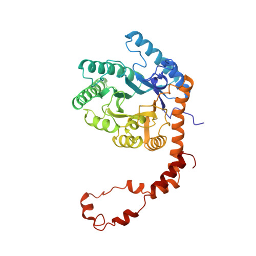
1XYL, 1XYM - PubMed Abstract:
The distinct roles of the two magnesium ions essential to the activity of D-xylose isomerase from Streptomyces olivochromogenes were examined. The enzyme-magnesium complex was isolated, and the stoichiometry of cation binding determined by neutron activation analysis to be 2 mol of magnesium per mole of enzyme. A plot of Mg2+ added versus Mg2+ bound to enzyme is consistent with apparent KD values of < or = 0.5-1.0 mM for one Mg2+ and < or = 2-5 mM for the second. A site-directed mutant of D-xylose isomerase was designed to remove the tighter, tetracoordinated magnesium binding site (site 1, Mg-1); Glu180 was replaced with Lys180. The stoichiometry of metal binding to this mutant, E180K, is 1 mol of magnesium per mole of enzyme. Ring-opening assays with 1-thioglucose (H2S released upon ring opening) show E180K catalyzes the opening of the sugar ring at 20% the rate of the wild-type, but E180K does not catalyze isomerization of glucose to fructose. Thus, the magnesium bound to Glu180 is essential for isomerization but not essential for ring opening. The X-ray crystallographic structures of E180K in the absence of magnesium and in the presence and absence of 250 mM glucose were obtained to 1.8-A resolution and refined to R factors of 17.7% and 19.7%, respectively. The wild-type and both E180K structures show no significant structural differences, except the epsilon-amino group of Lys180, which occupies the position usually occupied by the Mg-1.(ABSTRACT TRUNCATED AT 250 WORDS)
- Rosenstiel Basic Medical Sciences Research Center, Brandeis University, Waltham, Massachusetts 02254-9110.
Organizational Affiliation: