Crystal structure, at 2.6-A resolution, of the Streptomyces lividans xylanase A, a member of the F family of beta-1,4-D-glycanases.
Derewenda, U., Swenson, L., Green, R., Wei, Y., Morosoli, R., Shareck, F., Kluepfel, D., Derewenda, Z.S.(1994) J Biological Chem 269: 20811-20814
- PubMed: 8063693 Search on PubMed
- Primary Citation Related Structures:
1XAS - PubMed Abstract:
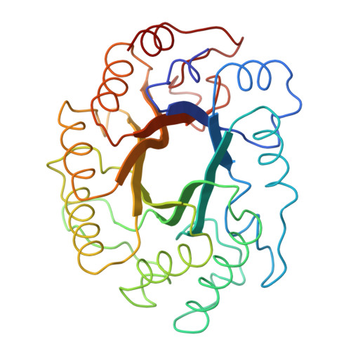
The crystal structure of the 32-kDa catalytic domain of the Streptomyces lividans xylanase A was solved by molecular isomorphous replacement methods and subsequently refined at 2.6-A resolution to a conventional crystallographic R factor of 0.21. This is the first successful structure determination of a member of the F family of endo-beta-1,4-D-glycanases. Unlike the recently determined xylanases of the G family (Wakarchuk, W. W., Campbell, R. L., Sung, W. L., Davoodi, J., and Yaguchi, M. (1994) Protein Sci. 3, 467-475), where the catalytic domains have a unique beta-sheet structure, the 32-kDa domain of the S. lividans xylanase A is folded into a complete (alpha/beta)8 barrel, the first such fold observed among beta-1,4-D-glycanases. The active site is located at the carbonyl end of the beta barrel. The crystal structure supports the earlier assignment of Glu-128 and Glu-236 as the catalytic amino acids (Moreau, A., Roberge, M., Manin, C., Shareck, F., Kluepfel, D., and Morosoli, R. (1994) Biochem. J., in press).
- Department of Biochemistry, University of Alberta, Edmonton, Canada.
Organizational Affiliation: