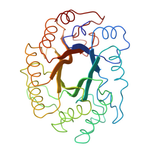
1XAS

CRYSTAL STRUCTURE, AT 2.6 ANGSTROMS RESOLUTION, OF THE STREPTOMYCES LIVIDANS XYLANASE A, A MEMBER OF THE F FAMILY OF BETA-1,4-D-GLYCANSES


Experimental Data Snapshot

  • Method: X-RAY DIFFRACTION
  • Resolution: 2.60 Å
  • R-Value Work: 0.210 
  • R-Value Observed: 0.210 

wwPDB Validation   3D Report Full Report


This is version 1.5 of the entry. See complete history


Literature

Crystal structure, at 2.6-A resolution, of the Streptomyces lividans xylanase A, a member of the F family of beta-1,4-D-glycanases.

Derewenda, U.Swenson, L.Green, R.Wei, Y.Morosoli, R.Shareck, F.Kluepfel, D.Derewenda, Z.S.

(1994) J Biol Chem 269: 20811-20814

  • Primary Citation of Related Structures:  
    1XAS

  • PubMed Abstract: 

    The crystal structure of the 32-kDa catalytic domain of the Streptomyces lividans xylanase A was solved by molecular isomorphous replacement methods and subsequently refined at 2.6-A resolution to a conventional crystallographic R factor of 0.21. This is the first successful structure determination of a member of the F family of endo-beta-1,4-D-glycanases. Unlike the recently determined xylanases of the G family (Wakarchuk, W. W., Campbell, R. L., Sung, W. L., Davoodi, J., and Yaguchi, M. (1994) Protein Sci. 3, 467-475), where the catalytic domains have a unique beta-sheet structure, the 32-kDa domain of the S. lividans xylanase A is folded into a complete (alpha/beta)8 barrel, the first such fold observed among beta-1,4-D-glycanases. The active site is located at the carbonyl end of the beta barrel. The crystal structure supports the earlier assignment of Glu-128 and Glu-236 as the catalytic amino acids (Moreau, A., Roberge, M., Manin, C., Shareck, F., Kluepfel, D., and Morosoli, R. (1994) Biochem. J., in press).


  • Organizational Affiliation

    Department of Biochemistry, University of Alberta, Edmonton, Canada.


Macromolecules
Find similar proteins by:  (by identity cutoff)  |  3D Structure
Entity ID: 1
MoleculeChains Sequence LengthOrganismDetailsImage
1,4-BETA-D-XYLAN XYLANOHYDROLASE299Streptomyces lividansMutation(s): 0 
EC: 3.2.1.8
UniProt
Find proteins for P26514 (Streptomyces lividans)
Explore P26514 
Go to UniProtKB:  P26514
Entity Groups  
Sequence Clusters30% Identity50% Identity70% Identity90% Identity95% Identity100% Identity
UniProt GroupP26514
Sequence Annotations
Expand
  • Reference Sequence
Experimental Data & Validation

Experimental Data

  • Method: X-RAY DIFFRACTION
  • Resolution: 2.60 Å
  • R-Value Work: 0.210 
  • R-Value Observed: 0.210 
  • Space Group: P 21 21 21
Unit Cell:
Length ( Å )Angle ( ˚ )
a = 69.52α = 90
b = 46.51β = 90
c = 85.5γ = 90
Software Package:
Software NamePurpose
X-PLORmodel building
PROLSQrefinement
X-PLORrefinement
X-PLORphasing

Structure Validation

View Full Validation Report


Entry History 

Deposition Data

Revision History  (Full details and data files)

  • Version 1.0: 1995-05-31
    Type: Initial release
  • Version 1.1: 2008-03-03
    Changes: Version format compliance
  • Version 1.2: 2011-07-13
    Changes: Version format compliance
  • Version 1.3: 2019-07-17
    Changes: Data collection, Other, Refinement description
  • Version 1.4: 2019-08-14
    Changes: Data collection, Refinement description
  • Version 1.5: 2024-02-14
    Changes: Data collection, Database references