Structural and Functional Analyses of an Archaeal XPF/Rad1/Mus81 Nuclease: Asymmetric DNA Binding and Cleavage Mechanisms
Nishino, T., Komori, K., Ishino, Y., Morikawa, K.(2005) Structure 13: 1183-1192
- PubMed: 16084390 Search on PubMed
- DOI: https://doi.org/10.1016/j.str.2005.04.024
- Primary Citation Related Structures:
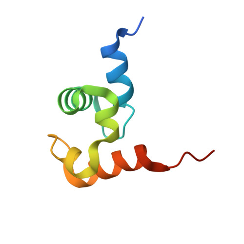
1X2I - PubMed Abstract:
XPF/Rad1/Mus81/Hef proteins recognize and cleave branched DNA structures. XPF and Rad1 proteins cleave the 5' side of nucleotide excision repair bubble, while Mus81 and Hef cleave similar sites of the nicked Holliday junction, fork, or flap structure. These proteins all function as dimers and consist of catalytic and helix-hairpin-helix DNA binding (HhH) domains. We have determined the crystal structure of the HhH domain of Pyrococcus furiosus Hef nuclease (HefHhH), which revealed the distinct mode of protein dimerization. Our structural and biochemical analyses also showed that each of the catalytic and HhH domains binds to distinct regions within the fork-structured DNA: each HhH domain from two separate subunits asymmetrically binds to the arm region, while the catalytic domain binds near the junction center. Upon binding to DNA, Hef nuclease disrupts base pairs near the cleavage site. It is most likely that this bipartite binding mode is conserved in the XPF/Rad1/Mus81 nuclease family.
- Department of Structural Biology, Biomolecular Engineering Research Institute, 6-2-3 Furuedai, Suita, Osaka 565-0874, Japan.
Organizational Affiliation: