Structure of the nondiscriminating aspartyl-tRNA synthetase from the crenarchaeon Sulfolobus tokodaii strain 7 reveals the recognition mechanism for two different tRNA anticodons
Sato, Y., Maeda, Y., Shimizu, S., Hossain, M.T., Ubukata, S., Suzuki, K., Sekiguchi, T., Takenaka, A.(2007) Acta Crystallogr D Biol Crystallogr 63: 1042-1047
- PubMed: 17881821 Search on PubMed
- DOI: https://doi.org/10.1107/S0907444907038292
- Primary Citation Related Structures:
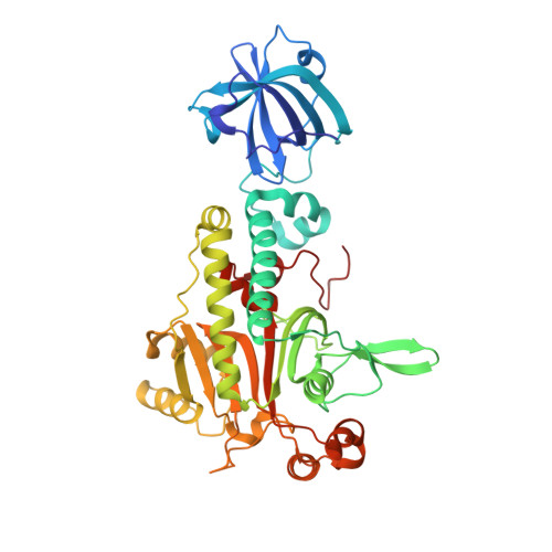
1WYD - PubMed Abstract:
In protein synthesis, 20 types of aminoacyl-tRNA synthetase (aaRS) are generally required in order to distinguish between the 20 types of amino acid so that each achieves strict recognition of the cognate amino acid and the cognate tRNA. In the crenarchaeon Sulfolobus tokodaii strain 7 (St), however, asparaginyl-tRNA synthetase (AsnRS) is missing. It is believed that AspRS instead produces Asp-tRNA(Asn) in addition to Asp-tRNA(Asp). In order to reveal the recognition mechanism for the two anticodons, GUC for aspartate and GUU for asparagine, the crystal structure of St-AspRS (nondiscriminating type) has been determined at 2.3 A resolution as the first example of the nondiscriminating type of AspRS from crenarchaea. A structural comparison with structures of discriminating AspRSs indicates that the structures are similar to each other overall and that the catalytic domain is highly conserved as expected. In the N-terminal domain, however, the binding site for the third anticodon nucleotide is modified to accept two pyrimidine bases, C and U, but not purine bases. The C base can bind to form a hydrogen bond to the surrounding main-chain amide group in the discriminating AspRS, while in the nondiscriminating AspRS the corresponding amino-acid residue is replaced by proline, which has no amide H atom for hydrogen-bond formation, thus allowing the U base to be accommodated in this site. In addition, the residues that cover the base plane are missing in the nondiscriminating AspRS. These amino-acid changes make it possible for both C and U to be accepted by the nondiscriminating AspRS. It is speculated that this type of nondiscriminating AspRS has been introduced into Thermus thermophilus through horizontal gene transfer.
- Graduate School of Bioscience and Biotechnology, Tokyo Institute of Technology, Nagatsuta, Midori-ku, Yokohama 226-8501, Japan.
Organizational Affiliation: